. 2022 Aug 30:1679:463389.
doi: 10.1016/j.chroma.2022.463389. Epub 2022 Jul 30.

Capillary electrophoresis Western blot using inkjet transfer to membrane

Affiliations

Capillary electrophoresis Western blot using inkjet transfer to membrane

Peter-Philip M Booth et al. J Chromatogr A. .

Abstract

Traditional Western blots are commonly used to separate and assay proteins; however, they have limitations including a long, cumbersome process and large sample requirements. Here, we describe a system for Western blotting where capillary gel electrophoresis is used to separate sodium dodecyl sulfate-protein complexes. The capillary outlet is threaded into a piezoelectric inkjetting head that deposits the separated proteins in a quasi-continuous stream of <100 pL droplets onto a moving membrane. Through separations at 400 V/cm and protein capture on a membrane moving at 2 mm/min, we are able to detect actin with a limit of detection at 8 pM, or an estimated 5 fg injected. Separation and membrane capture of sample containing 10 proteins ranging in molecular weights from 11 - 250 kDa was achieved in 15 min. The system was demonstrated with Western blots for actin, β-tubulin, ERK1/2, and STAT3 in human A431 epidermoid carcinoma cell lysate.

Keywords: CE-SDS; Western blot; immunoassay; inkjet; separation.


Conflict of interest statement

Declaration of competing interest The authors declare that they have no known competing financial interests or personal relationships that could have appeared to influence the work reported in this paper. The authors declare the following financial interests/personal relationships which may be considered as potential competing interests: Don T. Lamb reports financial support was provided by LI-COR Biosciences. Jon P. Anderson reports financial support was provided by LI-COR Biosciences. Michael D. Furtaw reports financial support was provided by LI-COR Biosciences. Michael D. Furtaw and Donald T. Lamb has patent Separation capillary inkjet dispensing with flat piezoelectric actuator pending to LI-COR, Inc. Michael D. Furtaw and Donald T. Lamb has patent Capillary electrophoresis inkjet dispensing issued to LI-COR, Inc.

Figures

Figure 1.
Figure 1.
a. In this system, samples are separated by CGE where the outlet end of the capillary is threaded into a piezoelectric inkjet head. Solutions are added by pipette into the sample reservoir. A pressure pump and a vacuum pump are connected to the sample reservoir and the inkjetting buffer allowing for pressure driven flow of solutions and the application of vacuum which is critical to inkjet operation. The inkjet dispenser is connected to both the inkjet buffer and the waste so that the buffer can flush and fill the dispenser and any excess buffer can be discarded to waste. A light-emitting diode (LED) and camera are in line with, but on opposite sides of the tip of the inkjetting dispenser to allow for stroboscope viewing of droplet formation. The cathode is in the sample reservoir while the anode is in the jetting buffer. Air connections are shown in yellow while fluidic connections are in blue, colored arrows indicate the direction of air/fluid flow. b. As proteins elute from the capillary, they are jetted onto a membrane on a moveable stage moving past the inkjet head at a constant rate. c. Zoomed-in view showing the tip of the piezoelectric inkjet head with the capillary placement. A single droplet is observed through stroboscope imaging.
Figure 2.
Figure 2.
A linear relationship is obtained between 1/migration time of each molecular weight marker and the log of its molecular weight for a NIR protein ladder with 10 proteins diluted 1:4000 in DI. Separations in a 10 cm capillary (40 μm I.D./150 μm O.D.) filled with sieving gel using a field strength of 400 V/cm (n = 3).
Figure 3.
Figure 3.
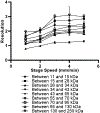
Peak heights in relative fluorescence units (RFU) for each molecular weight marker peaks measured at 400 V/cm across integer stage speeds from 1 – 5 mm/min using a 10 cm capillary (40 μm I.D./150 μm O.D.) filled with sieving gel. As stage speed increases, each peak spreads out, decreasing the signal height (plotted as mean ± SD for n = 3 replicates).
Figure 4.
Figure 4.
Peak heights in relative fluorescence units (RFU) of 70 kDa molecular weight marker measured at 300, 350, and 400 V/cm across integer stage speeds from 1 – 5 mm/min using a 10 cm capillary (40 μm I.D./150 μm O.D.) filled with sieving gel. The decrease in the number of inkjetted droplets deposited at any given point of the captured separation based on the stage speed correlates with the decrease in the observable signal (plotted as mean ± SD for n = 3 replicates).
Figure 5.
Figure 5.
Resolution between each adjacent molecular weight marker measured at 400 V/cm across integer stages speeds from 1 – 5 mm/min using a 10 cm capillary (40 μm I.D./150 μm O.D.) filled with sieving gel. Increasing stage speed led to increasing resolution until leveling off above 3 mm/min (plotted as mean ± SD for n = 3 replicates).
Figure 6.
Figure 6.
Resolution between the 34 and 43 kDa peaks at 300, 350 and 400 V/cm evaluated at integer stage speeds between 1 and 5 mm/min using a 10 cm capillary (40 μm I.D./150 μm O.D.) filled with sieving gel (plotted as mean ± SD for n = 3 replicates).
Figure 7.
Figure 7.
Peak heights in relative fluorescence units (RFU) of each NIR ladder protein measured for separations captured with the stage at normal operating height as well as ± 1 mm using a 10 cm capillary (40 μm I.D./150 μm O.D.) filled with sieving gel at 400 V/cm collect at a stage speed of 2 mm/min (plotted as mean ± SD for n = 3 replicates).
Figure 8.
Figure 8.
a. Immunoassay of decreasing concentrations of unlabeled actin were analyzed to determine the LOD of the system using a 10 cm capillary (40 μm I.D./150 μm O.D.) filled with sieving gel at 400 V/cm and collected at a stage speed of 2 mm/min. Direction of stage motion is such that droplets are collected from the bottom of the image to the top. At 9 pM, S/N was 5.2, giving an LOD of 8 pM. b. Electropherograms for 175 pM (dash-dotted line), 45 pM (dotted line), 35 pM (dashed line), and 9 pM (solid line). c. Electropherogram for just 9 pM actin.
Figure 9.
Figure 9.
Samples containing ladder and A431 human epidermoid carcinoma cell lysate were separated and probed for: a. actin (42 kDa), b. β-tubulin (55 kDa), c. STAT3 (79, 86 kDa), and d. ERK1/2 (42/44 kDa). Samples were separated in a 10 cm capillary (40 μm I.D./150 μm O.D.) filled with sieving gel at 400 V/cm. Separated samples were collected on membrane using a stage speed of 2 mm/min. Membrane scans show ladder proteins in red (scanned at 700 nm) while assayed proteins are in white (scanned at 800 nm). Electropherograms show ladder proteins (red) and assayed proteins (black). The first spot is the bromophenol blue dye in the A431 lysate.

Similar articles

Cited by

References

    1. Mishra M, Tiwari S, Gomes AV, Protein purification and analysis: next generation Western blotting techniques, Expert Rev. Proteom 14 (2017) 1037–1053, doi: 10.1080/14789450.2017.1388167. - DOI - PMC - PubMed
    1. He M, Herr AE, Microfluidic polyacrylamide gel electrophoresis with in situ immunoblotting for native protein analysis, Anal. Chem 81 (2009) 8177–8184, doi: 10.1021/ac901392u. - DOI - PubMed
    1. Hughes AJ, Herr AE, Microfluidic Western blotting, Proc. Natl. Acad. Sci 109 (2012) 21450–21455, doi: 10.1073/pnas.1207754110. - DOI - PMC - PubMed
    1. Gerver RE, Herr AE, Microfluidic western blotting of low-molecular-mass proteins, Anal. Chem 86 (2014) 10625–10632, doi: 10.1021/ac5024588. - DOI - PMC - PubMed
    1. Abdel-Sayed P, Yamauchi KA, Gerver RE, Herr AE, Fabrication of an open microfluidic device for immunoblotting, Anal. Chem (2017) 6. - PMC - PubMed

LinkOut - more resources