

Biochemical and biomechanical characterization of the cervical, thoracic, and lumbar facet joint cartilage in the Yucatan minipig
- PMID: 35933954
- PMCID: PMC9910803
- DOI: 10.1016/j.jbiomech.2022.111238
Biochemical and biomechanical characterization of the cervical, thoracic, and lumbar facet joint cartilage in the Yucatan minipig
Abstract
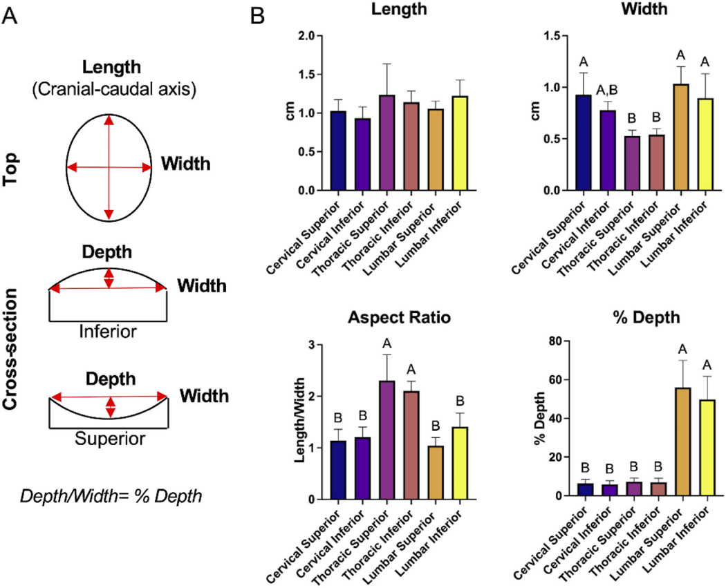
Facet joint arthrosis causes pain in approximately 7 % of the U.S. population, but current treatments are palliative. The objective of this study was to elucidate structure-function relationships and aid in the development of future treatments for the facet joint. This study characterized the articular surfaces of cervical, thoracic, and lumbar facet cartilage from skeletally mature (18-24 mo) Yucatan minipigs. The minipig was selected as the animal model because it is recognized by the U.S. Food and Drug Administration (FDA) and the American Society for Testing and Materials (ASTM) as a translationally relevant model for spine-related indications. It was found that the thoracic facets had a ∼2 times higher aspect ratio than lumbar and cervical facets. Lumbar facets had 6.9-9.6 times higher % depth than the cervical and thoracic facets. Aggregate modulus values ranged from 135 to 262 kPa, much lower than reported aggregate modulus in the human knee (reported to be 530-701 kPa). The tensile Young's modulus values ranged from 6.7 to 20.3 MPa, with the lumbar superior facet being 304 % and 286 % higher than the cervical inferior and thoracic superior facets, respectively. Moreover, 3D reconstructions of entire vertebral segments were generated. The results of this study imply that structure-function relationships in the facet cartilage are different from other joint cartilages because biochemical properties are analogous to other articular cartilage sources whereas mechanical properties are not. By providing functional properties and a 3D database of minipig facet geometries, this work may supply design criteria for future facet tissue engineering efforts.
Keywords: Biomechanics; Cartilage; Characterization; Facet joint; Minipig; Structure-function relationships; Zygapophyseal joint.
Copyright © 2022 Elsevier Ltd. All rights reserved.
Conflict of interest statement
Declaration of Competing Interest The authors declare the following financial interests/personal relationships which may be considered as potential competing interests: Drs. Athanasiou and Hu have an equity interest in Cartilage, Inc. Their relationship with Cartilage, Inc. has been reviewed and approved by the University of California, Irvine in accordance with its conflict-of-interest policies.
Figures


Similar articles
-
Biomechanical, biochemical, and histological characterization of sacroiliac joint cartilage in the Yucatan minipig.J Mech Behav Biomed Mater. 2024 Sep;157:106658. doi: 10.1016/j.jmbbm.2024.106658. Epub 2024 Jul 14. J Mech Behav Biomed Mater. 2024. PMID: 39018919 Free PMC article.
-
A Tribological Comparison of Facet Joint, Sacroiliac Joint, and Knee Cartilage in the Yucatan Minipig.Cartilage. 2021 Dec;13(2_suppl):346S-355S. doi: 10.1177/19476035211021906. Epub 2021 Jun 9. Cartilage. 2021. PMID: 34105385 Free PMC article.
-
Biochemical and biomechanical characterisation of equine cervical facet joint cartilage.Equine Vet J. 2018 Nov;50(6):800-808. doi: 10.1111/evj.12845. Epub 2018 May 17. Equine Vet J. 2018. PMID: 29658148
-
Spinal facet joint biomechanics and mechanotransduction in normal, injury and degenerative conditions.J Biomech Eng. 2011 Jul;133(7):071010. doi: 10.1115/1.4004493. J Biomech Eng. 2011. PMID: 21823749 Free PMC article. Review.
-
Facet Tropism in Lumbar Spine and Cervical Spine: A Systematic Review and Meta-Analysis.World Neurosurg. 2021 Mar;147:47-65. doi: 10.1016/j.wneu.2020.11.171. Epub 2020 Dec 9. World Neurosurg. 2021. PMID: 33309642
Cited by
-
Recent advancements in cartilage tissue engineering innovation and translation.Nat Rev Rheumatol. 2024 Jun;20(6):323-346. doi: 10.1038/s41584-024-01118-4. Epub 2024 May 13. Nat Rev Rheumatol. 2024. PMID: 38740860 Free PMC article. Review.
-
A cell bank paradigm for preclinical evaluation of an analogous cellular product for an allogeneic cell therapy.Biofabrication. 2024 Jun 3;16(3):10.1088/1758-5090/ad4de2. doi: 10.1088/1758-5090/ad4de2. Biofabrication. 2024. PMID: 38768586 Free PMC article.
-
The I-PREDICT 50th Percentile Male Warfighter Finite Element Model: Development and Validation of the Thoracolumbar Spine.Ann Biomed Eng. 2024 May 23. doi: 10.1007/s10439-024-03522-z. Online ahead of print. Ann Biomed Eng. 2024. PMID: 38780890
-
Tissue anisotropy and collagenomics in porcine penile tunica albuginea: Implications for penile structure-function relationships and tissue engineering.Acta Biomater. 2023 Oct 1;169:130-137. doi: 10.1016/j.actbio.2023.08.017. Epub 2023 Aug 13. Acta Biomater. 2023. PMID: 37579910 Free PMC article.
-
Biomechanical, biochemical, and histological characterization of sacroiliac joint cartilage in the Yucatan minipig.J Mech Behav Biomed Mater. 2024 Sep;157:106658. doi: 10.1016/j.jmbbm.2024.106658. Epub 2024 Jul 14. J Mech Behav Biomed Mater. 2024. PMID: 39018919 Free PMC article.
References
-
- ASTM, 2015. Standard Guide for Pre-clinical in vivo Evaluation of Spinal Fusion 1. Astm. 10.1520/F2884 - DOI
-
- Athanasiou KA, Agarwal A, Muffoletto A, Dzida FJ, Constantinides G, Clem M, 1995. Biomechanical properties of hip cartilage in experimental animal models. Clin. Orthop. Relat. Res 254–66. - PubMed
