. 2022 Oct;37(5):676-684.
doi: 10.1016/j.virs.2022.07.013. Epub 2022 Aug 5.

IFN-β1b induces OAS3 to inhibit EV71 via IFN-β1b/JAK/STAT1 pathway

Affiliations

IFN-β1b induces OAS3 to inhibit EV71 via IFN-β1b/JAK/STAT1 pathway

Baisong Zheng et al. Virol Sin. 2022 Oct.

Abstract

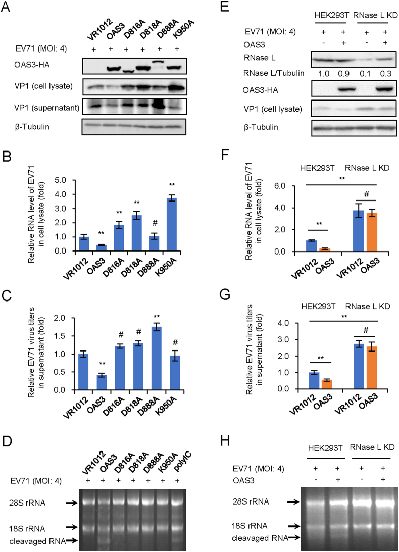
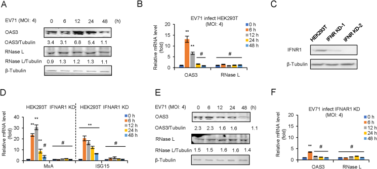
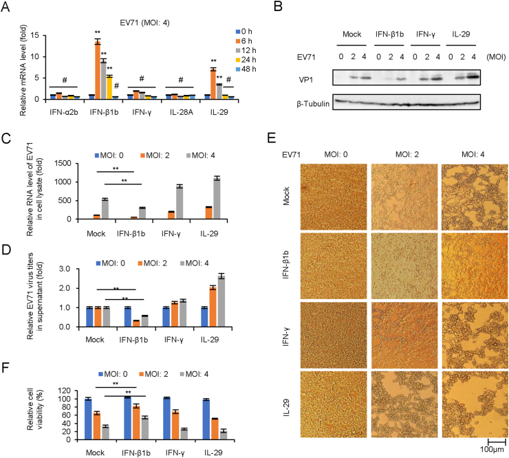
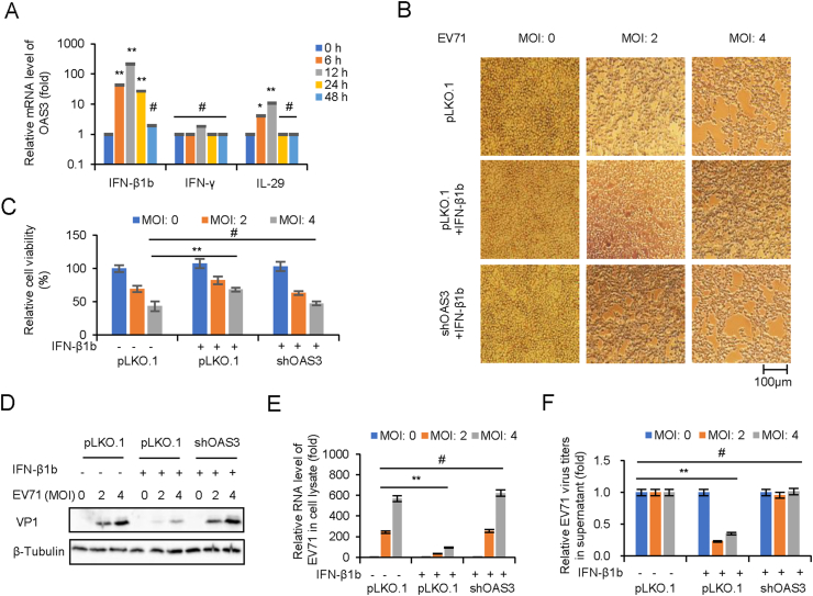
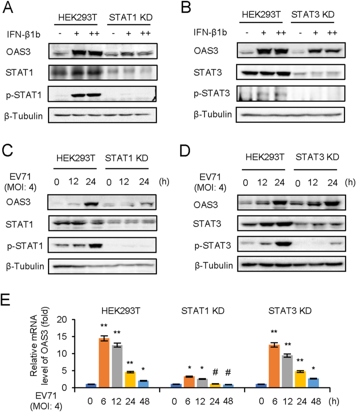
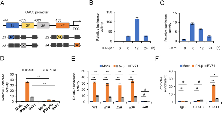
Enterovirus 71 (EV71) caused hand, foot and mouth disease (HFMD) is a serious threat to the health of young children. Although type I interferon (IFN-I) has been proven to control EV71 replication, the key downstream IFN-stimulated gene (ISG) remains to be clarified and investigated. Recently, we found that 2'-5'-oligoadenylate synthetases 3 (OAS3), as one of ISG of IFN-β1b, was antagonized by EV71 3C protein. Here, we confirm that OAS3 is the major determinant of IFN-β1b-mediated EV71 inhibition, which depends on the downstream constitutive RNase L activation. 2'-5'-oligoadenylate (2-5A) synthesis activity deficient mutations of OAS3 D816A, D818A, D888A, and K950A lost resistance to EV71 because they could not activate downstream RNase L. Further investigation proved that EV71 infection induced OAS3 but not RNase L expression by IFN pathway. Mechanically, EV71 or IFN-β1b-induced phosphorylation of STAT1, but not STAT3, initiated the transcription of OAS3 by directly binding to the OAS3 promoter. Our works elucidate the immune regulatory mechanism of the host OAS3/RNase L system against EV71 replication.

Keywords: 2′-5′-oligoadenylate synthetases 3 (OAS3); Enterovirus 71 (EV71); IFN-β1b; JAK/STAT; RNase L.


Conflict of interest statement

Conflict of interest The authors declare that they have no conflict of interest.

Figures

Fig. 1
Fig. 1
OAS3 inhibits EV71 replication depending on RNase L activation. 2-5A synthesis activity of OAS3 determines the inhibitory effect on EV71 replication. A WT and inactivated mutants of OAS3 transfected HEK293T cells were infected with EV71 at an MOI of 4. The expression levels of exogenous OAS3 and VP1 were examined by Western blotting. B Viral RNA in cell lysate was examined by RT-qPCR. The RNA level of VR1012 was set as “1”. C Viral titers in the supernatants were determined by the plaque assay. D RNase L activation was examined by agarose electrophoresis of total rRNA of cells. E The negative control pLKO.1 and RNase L knockdown (RNase L KD) HEK293T cells were transfected with OAS3-expressing or control plasmids. Cells were infected with EV71 at an MOI of 4. The expression levels of endogenous RNase L, exogenous OAS3, and VP1 were examined by Western blotting. F Viral RNA in cell lysate was examined by RT-qPCR. The RNA level of VR1012 was set as “1”. G Viral titers in the supernatants were determined by the plaque assay. H RNase L activation was examined by agarose electrophoresis of total rRNA of cells. The results represent the means ​± ​standard deviation from three independent experiments. Statistical significance was analyzed using Student's t-test (# no significance, ∗∗P ​< ​0.01).
Fig. 2
Fig. 2
EV71 infection induces OAS3 but not RNase L expression by IFN pathway. EV71 infection upregulates the expression of OAS3 but not RNase L. HEK293T cells were infected with EV71 at an MOI of 4, and the expression levels of OAS3 and RNase L were examined by Western blotting (A). The mRNA levels of OAS3 and RNase L were examined by RT-qPCR (B). The RNA level of “0 ​h” was set as “1”. C Construction of IFNAR1 CRISPR/Cas9 knockdown cell line. The efficiency of knockdown was examined by Western blotting. D The negative control pLKO.1 and IFNAR1 knockdown (IFNAR1 KD) HEK293T cells were treated with 250 U/mL IFN-β1b for 48 ​h, and the expression levels of MxA and ISG15 were examined by RT-qPCR at different time points. The RNA level of “0 ​h” was set as “1”. E IFNAR1 KD HEK293T cells were infected with EV71 at an MOI of 4, and the expression levels of OAS3 and RNase L were examined by Western blotting. F The mRNA levels of OAS3 and RNase L were examined by RT-qPCR. The RNA level of “0 ​h” was set as “1”. The results represent the means ​± ​standard deviation from three independent experiments. Statistical significance was analyzed using Student's t-test (# no significance, ∗∗P ​< ​0.01).
Fig. 3
Fig. 3
IFN-β1b significantly inhibits EV71 replication and viral cytopathic effect. A EV71 infection induces the expression of IFN-β1b in HEK293T cells. HEK293T cells were infected with EV71 at an MOI of 4, and the expression levels of IFNs were examined by RT-qPCR at different time points. The RNA level of “0 ​h” was set as “1”. B IFN-β1b plays an antiviral effect in EV71 infected HEK293T cells. After 250 U/mL IFN-β1b, 250 U/mL IFN-γ, and 250 U/mL IL29 treated HEK293T cells for 24 ​h, cells were infected EV71 at different MOI. The expression levels of VP1 were examined by Western blotting. C The expression levels of EV71 viral RNA were examined by RT-qPCR. The RNA level at an MOI of 0 was set as “1”. D Viral titers in the supernatants were determined by the plaque assay. E Apparent CPE was observed at 48 ​h.p.i. F Cell viability in the experiment of Fig. 3E was measured by CCK8. The cell viability at an MOI of 0 was set as “1”. The results represent the means ​± ​standard deviation from three independent experiments. Statistical significance was analyzed using Student's t-test (# no significance, ∗∗P ​< ​0.01).
Fig. 4
Fig. 4
OAS3 plays an important role in IFN-β1b-mediated inhibition of EV71. A IFN-β treatment induces the expression of OAS in HEK293T cells. HEK293T cells were treated with 250 U/mL IFN-β1b, 250 U/mL IFN-γ, and 250 U/mL IL-29 for 48 ​h, and the expression levels of OAS3 were examined by RT-qPCR at different time points. The RNA level of “0 ​h” was set as “1”. B HEK293T shRNA control (pLKO.1) and shOAS3 cells were pretreated with or without 250 U/mL IFN-β for 24 ​h prior to EV71 infection at MOIs of 2 and 4. Apparent CPE was observed at 48 ​h.p.i. C Cell viability in the experiment of Fig. 4B was measured by CCK8. D The expression levels of VP1 were examined by Western blotting. E The expression levels of EV71 viral RNA were examined by RT-qPCR. The RNA level at an MOI of 0 was set as “1”. F Viral titers in the supernatants were determined by the plaque assay. The results represent the means ​± ​standard deviation from three independent experiments. Statistical significance was analyzed using Student's t-test (# no significance, ∗∗P ​< ​0.01).
Fig. 5
Fig. 5
IFN-β1b-mediated OAS3 induction depends on STAT1 phosphorylation. A The negative control pLKO.1 and STAT1 knockdown (KD) HEK293T cells were treated with 125 U/mL (+) and 250 U/mL (++) IFN-β1b for 24 ​h as indicated, and the expression of endogenous OAS3, STAT1, and p-STAT1 were examined by Western blotting. B The negative control pLKO.1 and STAT3 KD HEK293T cells were treated with 125 U/mL (+) and 250 U/mL (++) IFN-β1b for 24 ​h as indicated, and the expression of endogenous OAS3, STAT1, and p-STAT1 were examined by Western blotting. C The negative control pLKO.1 and STAT1 KD HEK293T cells were infected with EV71 at an MOI of 4, and the expression of endogenous OAS3, STAT1, and p-STAT1 were examined by Western blotting at different time points. D The negative control pLKO.1 and STAT3 KD HEK293T cells were infected with EV71 at an MOI of 4, and the expression of endogenous OAS3, STAT1, and p-STAT1 were examined by Western blotting at different time points. E The negative control pLKO.1, STAT1 KD, and STAT3 KD HEK293T cells were infected with EV71 at an MOI of 4, and the expression levels of OAS3 were examined by RT-qPCR at different time points. The RNA level of “0 ​h” was set as “1”. The results represent the means ​± ​standard deviation from three independent experiments. Statistical significance was analyzed using Student's t-test (# no significance, ∗P ​< ​0.05, ∗∗P ​< ​0.01).
Fig. 6
Fig. 6
STAT1 binds to the OAS3 promoter. A Schematic illustration showing four potential STAT1 binding sites in the OAS3 promoter region. B, C HEK293T cells were transfected with firely luciferase reporter harboring wild type (WT) promoter regions of OAS3 together with Renilla luciferase reporter for 12 ​h. Cells were treated with 250 U/mL IFN-β1b or infected with EV71 at an MOI of 4 for 24 ​h, and dual-luciferase reporter assay was measured at different time points. Relative luciferase activity of “0 ​h” was set as “1”. D The negative control pLKO.1 and STAT1 knockdown (KD) HEK293T cells were transfected with firefly luciferase reporter harboring WT promoter regions of OAS3 together with Renilla luciferase reporter for 12 ​h. Cells were treated with 250 U/mL IFN-β1b or infected with EV71 at an MOI of 4 and dual-luciferase reporter assay was measured at 24 ​h. Relative luciferase activity of “Mock” was set as “1”. E HEK293T cells were transfected with firefly luciferase reporter harboring WT or mutant promoter regions of OAS3 together with Renilla luciferase reporter for 12 ​h. Cells were treated with 250 U/mL IFN-β1b or infected with EV71 at an MOI of 4 and dual-luciferase reporter assay was measured at 24 ​h. Relative luciferase activity of “Mock” was set as “1”. F HEK293T cells were treated with 250 U/mL IFN-β1b or infected with EV71 at an MOI of 4 for 24 ​h. A chromatin immunoprecipitation assay was performed to confirm the binding of STAT1 on OAS3 promoter. The RNA level of “Mock” was set as “1”. The results represent the means ​± ​standard deviation from three independent experiments. Statistical significance was analyzed using Student's t-test (# no significance, ∗P ​< ​0.05, ∗∗P ​< ​0.01).

Similar articles

Cited by

References

    1. Birdwell L.D., Zalinger Z.B., Li Y., Wright P.W., Elliott R., Rose K.M., Silverman R.H., Weiss S.R. Activation of rnase l by murine coronavirus in myeloid cells is dependent on basal oas gene expression and independent of virus-induced interferon. J. Virol. 2016;90:3160–3172. - PMC - PubMed
    1. Chebath J., Benech P., Revel M., Vigneron M. Constitutive expression of (2'-5') oligo a synthetase confers resistance to picornavirus infection. Nature. 1987;330:587–588. - PubMed
    1. Choi U.Y., Kang J.S., Hwang Y.S., Kim Y.J. Oligoadenylate synthase-like (oasl) proteins: dual functions and associations with diseases. Exp. Mol. Med. 2015;47:e144. - PMC - PubMed
    1. Coccia E.M., Romeo G., Nissim A., Marziali G., Albertini R., Affabris E., Battistini A., Fiorucci G., Orsatti R., Rossi G.B., et al. A full-length murine 2-5a synthetase cdna transfected in nih-3t3 cells impairs emcv but not vsv replication. Virology. 1990;179:228–233. - PubMed
    1. Floyd-Smith G. (2'-5')an-dependent endoribonuclease: enzyme levels are regulated by ifn beta, ifn gamma, and cell culture conditions. J. Cell. Biochem. 1988;38:13–21. - PubMed