. 2022 Oct:26:88-96.
doi: 10.1016/j.jtos.2022.07.012. Epub 2022 Aug 5.

Benzalkonium chloride, a common ophthalmic preservative, compromises rat corneal cold sensitive nerve activity

Affiliations

Benzalkonium chloride, a common ophthalmic preservative, compromises rat corneal cold sensitive nerve activity

Evguenia Ivakhnitskaia et al. Ocul Surf. 2022 Oct.

Abstract

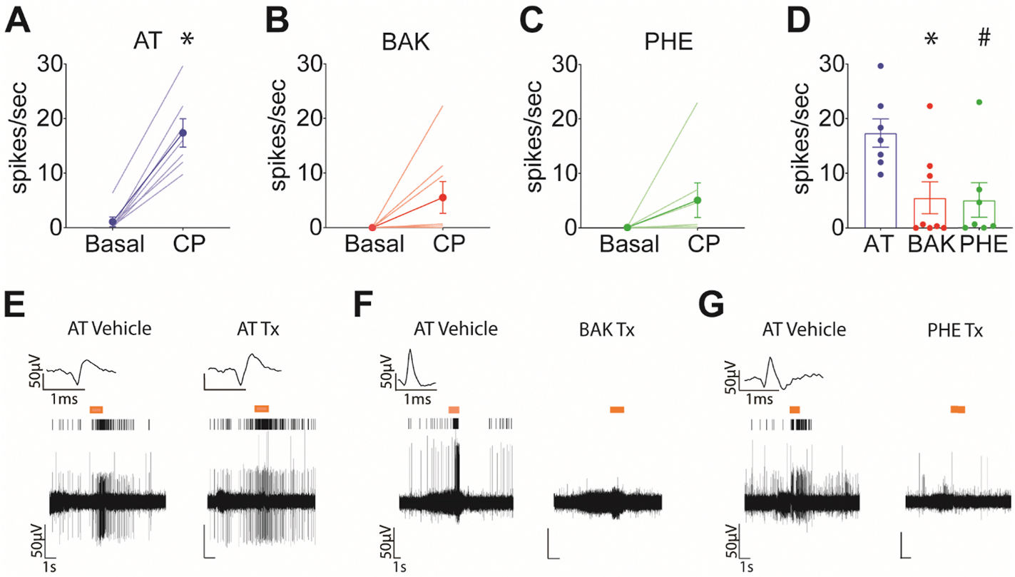
Purpose: Corneal nerves comprise the densest sensory network in the body. Dysfunction of the corneal cold sensitive neurons (CSN) is implicated in ophthalmic disorders, including Dry Eye Disease, the most common ocular surface disorder. The preservative Benzalkonium chloride (BAK) and the mydriatic agent Phenylephrine hydrochloride (PHE) are considered to be inactive at the level of the CSNs. The purpose of this study is to test the impacts of continuous exposures to BAK or PHE at their clinically used concentrations on corneal nerve structure and function.

Methods: In vivo extracellular electrophysiology of the rat trigeminal ganglion was used to monitor CSN functional response to stimuli mimicking physiological states and stressors of the cornea. Corneal nerve structure was evaluated by immunostaining.

Results: Among the tested stimuli, cold probe receptive field stimulation and hyperosmolar stress were the most sensitive methods of detecting activity changes. CSN activity was attenuated after 30 min exposure to either PHE or BAK. After an hour-long washout period, BAK-treated neurons failed to recover activity while PHE-treated neurons showed signs of functional recovery. Intraepithelial nerve density was reduced and nerve fragmentation was increased in BAK-treated corneas, while PHE exposure left corneal nerves structurally intact.

Conclusions: Our study suggests that prolonged ocular instillations of BAK or PHE alter CSN activity through two different processes - irreversible neuronal damage in the case of BAK vs. reversible attenuation in the case of PHE.

Keywords: Benzalkonium chloride; Cold sensitive nerves; Cornea; In vivo electrophysiology; Nerve damage; Nerve function; Nerve structure; Sensation.


Conflict of interest statement

Declaration of competing interest The authors have no conflicts to disclose relevant to the production and publication of this work.

Figures

Fig. 1.
Fig. 1.
Experimental approach and characterization profiles of corneal cold sensitive neurons. A, Illustration of the recording arrangement and characterization protocol on the ocular surface. A cranial window was created in order to advance a tungsten microelectrode through the brain into the trigeminal ganglion. Once a putative corneal neuron was located in the ganglion, the ocular surface was stimulated with a number of characterization stimuli. Solution-based stimuli were applied in an ocular well. B, A sample trace of a CSN average spiking activity during characterization. During periods of “dryness,” the receptive field of the neurons was probed with an ice-cooled metal probe (blue arrow) and von Frey filaments (green arrow). During instillations of AT on the ocular surface, the AT temperature was altered to determine the neuron’s temperature activation thresholds. A 600 mOsm AT solution was applied to test the neuron’s sensitivity to hyperosmolar solutions. C, Activity trace of a LT-CSN activated by a temperature change of less than 1 °C during a ramp to 9 °C. Shown activity traces are characterizations following a period of 30 min AT instillation to establish baseline; orange dotted line represents the approximate temperature of activation. D, Example of a HT-CSN activated by ocular surface cooling of greater than 2.5 °C. E, Example of a UHT-CSN activated at cooling temperatures at least 8 °C cooler than holding temperature. F, Experimental design. Initial characterization to determine neuronal subtype was followed by 30 min AT instillation to establish baseline responsiveness by assessing neuronal properties. Then neurons were exposed to one of three differential treatments for 30 min and responses were re-assessed. An hour long AT washout step was administered and the neuronal properties were re-assessed a final time to test the reversibility of treatment effects on activity.
Fig. 2.
Fig. 2.
Baseline responsiveness of corneal cold sensitive neurons to cold probe application and hyperosmolar stress. A, Cold probe (CP) stimulation induced a significant increase in firing of vehicle-assigned neurons at baseline, prior to application of AT vehicle treatment (Tx) (Wilcoxon test, W(7) = 28, p = 0.0156). B, Stimulus effect of CP application at baseline of neurons assigned to BAK Tx (W(8) = 36, p = 0.0078). C, Similarly, a stimulus effect of CP application was observed at baseline for neurons assigned to PHE Tx (W(7) = 28, p = 0.0156). D, Bar graph highlighting that the average response to CP stimulus did not vary at baseline among neurons assigned to the three Tx groups (Kruskal Wallis test, H(2,19) = 0.4678, p = 0.8024). E, At baseline ocular surface instillation with Hyperosmolar (HOsm) solution did not induce a significant stimulus effect for the AT vehicle-assigned group of neurons (W(6) = 11, p = 0.3125). F, The stimulus effect of HOsm instillation in the BAK-assigned group trended towards significance at baseline (W(6) = 19, p = 0.0625). G, HOsm stress induced a significant stimulus effect at baseline in the PHE-assigned group of neurons (W(6) = 21, p = 0.0313). H, Bar graph highlighting that the average response during HOsm instillation did not differ at baseline among neurons assigned to differential treatments (H(2,15) = 0.2456, p = 0.8968).
Fig. 3.
Fig. 3.
BAK and PHE treatments attenuate CSN response to cold probe stimulation. A, AT treated neurons showed a significant stimulus effect of cold probe stimulation (Wilcoxon test, W(7) = 28, p = 0.0156). B, There was a trend but no longer a significant effect of CP stimulation for 0.01% BAK-treated neurons (W(8) = 27, p = 0.0625). C, Similarly, 2.5% PHE-treated neurons showed a trend but lacked a significant response to cold probe stimulation (W(7) = 23, p = 0.0625). D, Bar graph showcasing significantly diminished firing to CP stimulation after BAK or PHE Tx compared to vehicle-treated neurons (Kruskal Wallis test, H(2,19) = 8.253, *p = 0.0111; p = 0.0201 AT vs BAK, #p = 0.0309 AT vs PHE by Dunn’s multiple comparison test). E, Activity trace of a representative neuron stimulated with CP at baseline and following 30 min Tx with AT vehicle. F, Activity trace of a representative neuron stimulated with CP at baseline and following 30 min Tx with BAK. G, Activity trace of a representative neuron stimulated with CP at baseline and following 30 min Tx with PHE. Insets display the waveform shape and amplitude of the neuron of interest; absence of inset signifies disappearance of waveform of interest at the tested time point. Activity of the cell of interest is plotted using a line raster plot. Orange bars represent delivery of cold probe stimulation. Stimulation artifact has been removed from the traces for clarity.
Fig. 4.
Fig. 4.
BAK treatment attenuates CSN response to hyperosmolar stress. A, AT treated neurons showed a significant stimulus effect of hyperosmolar stress (Wilcoxon test, W(7) = 28, p = 0.0156). B, BAK-treated neurons did not respond to Hosm stimulus (W(7) = 2, p = 0.8750). C, PHE-treated neurons showed a trend but lacked a significant response to HOsm stimulus (W(6) = 19, p = 0.0625). D, Neurons exposed to BAK fired at a significantly lower rate during HOsm stimulus compared to AT-treated neurons (Kruskal Wallis H(2,17)=14.50, p < 0.0001, p = 0.0003 AT vs BAK, p = 0.0821 AT vs PHE by Dunn’s test). E, Activity trace of a representative neuron during a HOsm instillation at baseline and following 30 min Tx with AT vehicle. F, Activity trace of a representative neuron during a HOsm instillation at baseline and following 30 min Tx with 0.01% BAK. G, Activity trace of a representative neuron during a HOsm instillation at baseline and following 30 min Tx with 2.5% PHE. Insets display the waveform shape and amplitude of the neuron of interest; absence of inset signifies disappearance of waveform of interest at the tested time point. Activity of the neuron of interest is plotted using a line raster plot. Orange bars represent the period of HOsm instillation on the ocular surface. Stimulation artifact has been removed from the traces for clarity.
Fig. 5.
Fig. 5.
BAK-induced attenuation of CSN response to cold probe stimulus is irreversible in contrast to the reversible effects of PHE treatment. A, As expected, after washout AT treated neurons retained a significant response to CP stimulation (Wilcoxon test, W(6) = 21, p = 0.0313). B, BAK treated neurons failed to recover responsiveness to cold probe stimulation even after an hour-long washout period (W(8) = 16, p = 0.25). C, In contrast, the washout period was sufficient to restore responsiveness to CP of previously PHE-treated neurons (W(7) = 28, p = 0.0156). D, Bar graph illustrating that firing rates to CP stimulation of BAK-treated neurons remained significantly reduced after the washout period, while firing rates of PHE-treated neurons were indistinguishable compared to AT-treated neurons (Kruskal-Wallis test, H(2,18) = 10.40, p = 0.0022; **p = 0.0086 AT vs BAK, p > 0.9999 AT vs PHE by Dunn’s test). E, Activity traces of a representative neuron responding to CP stress following vehicle treatment and washout. F, Activity traces of a representative neuron responding to CP stress following 0.01% BAK treatment and washout. G, Activity traces of a representative neuron responding to CP stress following 2.5% PHE treatment and washout. Insets display the waveform shape and amplitude of the neuron of interest; absence of inset signifies disappearance of waveform of interest at the tested time point. Activity of the neuron of interest is plotted using a line raster plot. Orange bars represent delivery of cold probe stimulation. Stimulation artifact has been removed from the traces for clarity.
Fig. 6.
Fig. 6.
BAK-induced attenuation of CSN response to hyperosmolar stress is irreversible. A, After washout, AT-treated neurons continued to respond to HOsm stress stimulus (Wilcoxon test, W(6) = 21, p = 0.0313). B, In contrast, BAK-treated neurons failed to recover responsiveness to HOsm stress (W(8) = 12, p = 0.5000). C, Conversely, PHE treated neurons recovered responsiveness to HOsm stress after washout (W(7) = 26, p = 0.0313). D, Bar graph summary of CSN activity during HOsm instillation following washout of AT, BAK, and PHE treatments (Kruskal-Wallis test, H(2,18) = 11.06, p = 0.0013; **p = 0.0021 AT vs BAK, p = 0.3562 AT vs PHE by Dunn’s test). E, Activity traces of a representative neuron responding to HOsm stress following vehicle treatment and washout. F, Activity traces of a representative neuron responding to HOsm stress following 0.01% BAK treatment and washout. G, Activity traces of a representative neuron responding to HOsm stress following 2.5% PHE treatment and washout. Insets display the waveform shape and amplitude of the neuron of interest; absence of inset signifies disappearance of waveform of interest at the tested time point. Activity of the neuron of interest is plotted using a line raster plot. Orange bars represent period of HOsm instillation on the ocular surface. Stimulation artifact has been removed from the traces for clarity.
Fig. 7.
Fig. 7.
Corneas treated with BAK show delayed signs of structural compromise. A, Subbasal corneal nerve architecture in the central cornea of rats that were sacrificed immediately after a 30 m ocular instillation with AT (left), 0.01% BAK in AT (middle) or 2.5% PHE in AT (right). Inset denotes the magnified view of a representative region (smaller rectangle) of corneal innervation. B, Subbasal corneal nerve architecture in the central cornea of rats that were sacrificed after an hour-long AT washout following a 30 min ocular instillation with AT (left), 0.01% BAK in AT (middle), and 2.5% PHE in AT (right). C, Corneal nerve density did not differ among treatments in rats sacrificed immediately post 30 m treatment (One-way ANOVA F(2, 19) = 0.5590, p = 0.5809). D, Similarly, there was no significant difference in nerve fragmentation of corneas collected immediately after 30 m treatment (Brown-Forsythe ANOVA F*(2.000, 5.823) = 2.655, p = 0.1515). E, Corneas collected from BAK-treated rats following an hour long washout period displayed significantly reduced corneal nerve density (F(2, 17) = 8.314, p = 0.0030, followed by Dunnett’s multiple comparisons test with **p = 0.0024 BAK + wash vs AT + wash, p = 0.9939 BAK + wash vs PHE + wash). F, Similarly, fragmentation of corneal nerves was significantly increased only in the BAK-treated corneas collected after an hour long washout; (F(2, 17) = 36.54, p < 0.0001, ****p < 0.0001 AT + wash vs BAK + wash, p = 0.8758 AT + wash vs PHE + wash). Nerve fragments/area indicates the number of nerve fragments within a 0.001 mm2 nerve area. Each point represents one cornea. Scale bars = 200 μm.

Similar articles

Cited by

References

    1. Rózsa AJ, Beuerman RW. Density and organization of free nerve endings in the corneal epithelium of the rabbit. Pain 1982;14(2):105–20. - PubMed
    1. Marfurt CF, Kingsley RE, Echtenkamp SE. Sensory and sympathetic innervation of the mammalian cornea. A retrograde tracing study. Investigative Ophthalmology & Visual Science 1989;30(3):461–72. - PubMed
    1. Belmonte C, et al. TFOS DEWS II pain and sensation report. Ocul Surf 2017;15(3):404–37. - PMC - PubMed
    1. Sacchetti M, Lambiase A. Neurotrophic factors and corneal nerve regeneration. Neural Regen Res 2017;12(8):1220–4. - PMC - PubMed
    1. Shaheen B, Bakir M, Jain S. Corneal nerves in health and disease. Survey of ophthalmology 2014;59(3):263–85. - PMC - PubMed

Publication types

Substances

LinkOut - more resources