. 2022 Sep;36(9):653-675.
doi: 10.1007/s10822-022-00470-5. Epub 2022 Aug 8.

Unveiling the G4-PAMAM capacity to bind and protect Ang-(1-7) bioactive peptide by molecular dynamics simulations

Affiliations

Unveiling the G4-PAMAM capacity to bind and protect Ang-(1-7) bioactive peptide by molecular dynamics simulations

L América Chi et al. J Comput Aided Mol Des. 2022 Sep.

Abstract

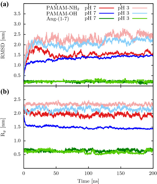
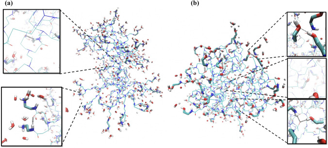
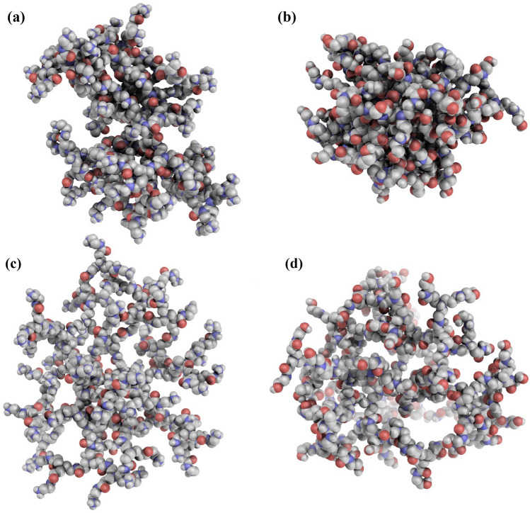
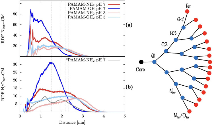
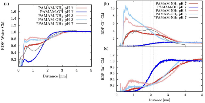
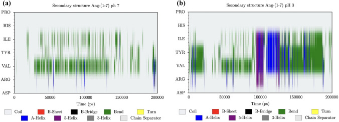
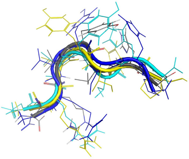
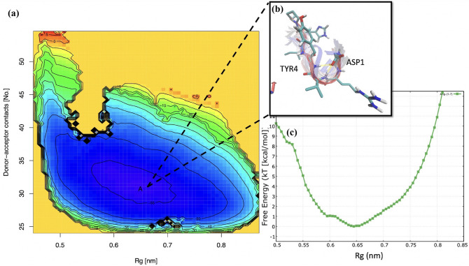
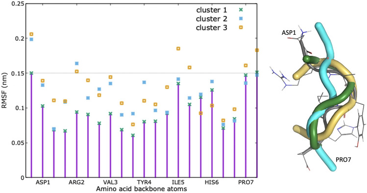
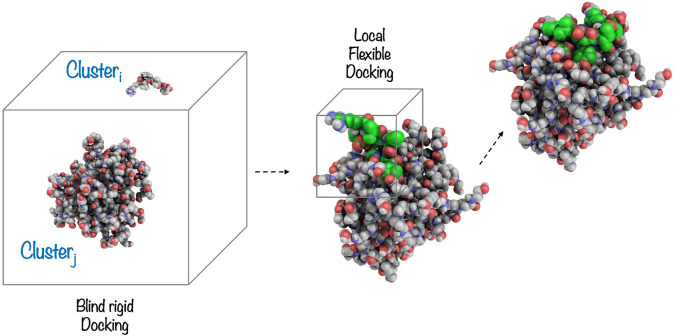
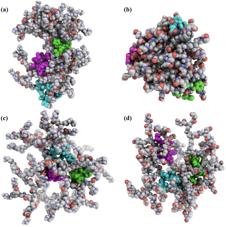
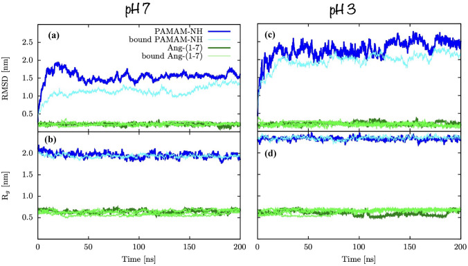
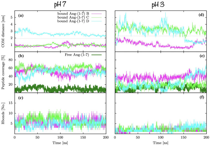
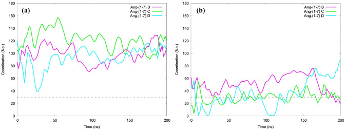
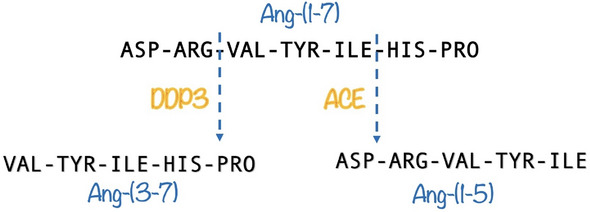
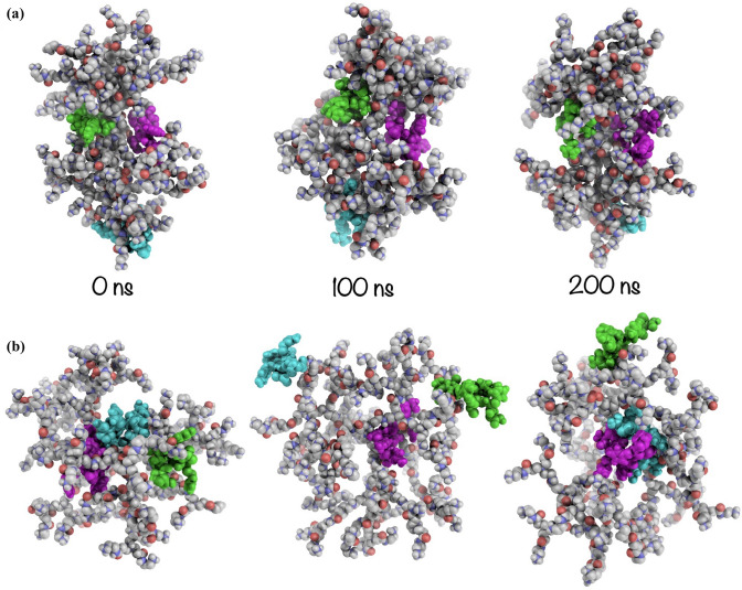
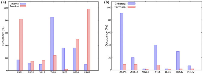
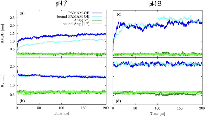
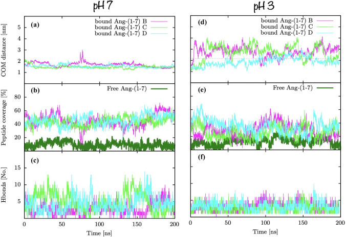
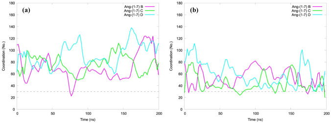
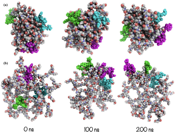
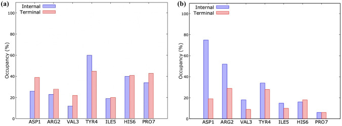
Angiotensin-(1-7) re-balance the Renin-Angiotensin system affected during several pathologies, including the new COVID-19; cardiovascular diseases; and cancer. However, one of the limiting factors for its therapeutic use is its short half-life, which might be overcome with the use of dendrimers as nanoprotectors. In this work, we addressed the following issues: (1) the capacity of our computational protocol to reproduce the experimental structural features of the (hydroxyl/amino)-terminated PAMAM dendrimers as well as the Angiotensin-(1-7) peptide; (2) the coupling of Angiotensin-(1-7) to (hydroxyl/amino)-terminated PAMAM dendrimers in order to gain insight into the structural basis of its molecular binding; (3) the capacity of the dendrimers to protect Angiotensin-(1-7); and (4) the effect of pH changes on the peptide binding and covering. Our Molecular-Dynamics/Metadynamics-based computational protocol well modeled the structural experimental features reported in the literature and our double-docking approach was able to provide reasonable initial structures for stable complexes. At neutral pH, PAMAM dendrimers with both terminal types were able to interact stably with 3 Angiotensin-(1-7) peptides through ASP1, TYR4 and PRO7 key amino acids. In general, they bind on the surface in the case of the hydroxyl-terminated compact dendrimer and in the internal zone in the case of the amino-terminated open dendrimer. At acidic pH, PAMAM dendrimers with both terminal groups are still able to interact with peptides either internalized or in its periphery, however, the number of contacts, the percentage of coverage and the number of hydrogen bonds are lesser than at neutral pH, suggesting a state for peptide release. In summary, amino-terminated PAMAM dendrimer showed slightly better features to bind, load and protect Angiotensin-(1-7) peptides.

Keywords: Ang-(1-7); Dendrimer; G4-PAMAM; Metadynamics; Molecular dynamics; Nanocarrier; PAMAM-OH; Peptide.


Conflict of interest statement

The authors have no competing interests to declare that are relevant to the content of this article.

Figures

Fig. 1
Fig. 1
Diagram representing the main metabolic effects driven by the Renin Angiotensin System bioactive peptides (Ang II and Ang-(1-7)) when interacting with its cell receptors AT1 and MAS, respectively. The main proteolytic cleavage of Ang-(1-7) by angiotensin-converting enzyme (ACE) and dipeptidyl peptidase 3 (DPP3), which is associated with Ang-(1-7) rapid degradation and limited therapeutic application
Fig. 2
Fig. 2
Structural features of PAMAM-NH2, PAMAM-OH and Ang-(1-7) systems. a RMSD and b Rg values as function of time
Fig. 3
Fig. 3
Waters sorrounding group terminals and hydrogen bonds formed between terminal groups and waters/dendrimer internal groups. a PAMAM-NH2 and b PAMAM-OH, at neutral pH
Fig. 4
Fig. 4
Dendrimers shape comparision. a PAMAM-NH2n, b PAMAM-OHn, c PAMAM-NH2a and d PAMAM-OHa at 200 ns of MD simulation. Leters “a” and “n” states for neutral and acidic pH
Fig. 5
Fig. 5
Dendrimers cavities comparision. a PAMAM-NH2n, b PAMAM-OHn, c PAMAM-NH2a and d PAMAM-OHa at 200 ns. Leters “a” and “n” states for neutral and acidic pH
Fig. 6
Fig. 6
Radial distribution function of molecular groups with respect of the dendrimer COM. a Internal nitrogen atoms and b terminal nitrogen/oxygen atoms. Schematic diagram at the right of the panel showing the internal and terminal nitrogens. GX states for the dendrimer generation. *Thinner grey line states for comparison from previous work in the literature [31]. Grey vertical dashed line represents the Rg of the dendrimers
Fig. 7
Fig. 7
Radial distribution function of molecules with respect of the dendrimer COM. a Water molecules and b Cl- ions and c Na+ ions. *Thinner grey line states for comparison from previous work in the literature [31]. Grey vertical dashed line represents the Rg of the dendrimers
Fig. 8
Fig. 8
Ang-(1-7) secondary structure as a function of time. a At neutral pH and b at acidic pH
Fig. 9
Fig. 9
Superimposition of: experimental NMR structure obtained at acidic pH (grey), structure repeated 40 % of the frames in classical MD simulation performed under neutral pH (blue), structure repeated 35 % of the frames in metadynamics simulation performed under neutral pH (cyan), structure repeated 60 % of the frames in classical MD simulation performed under acidic pH (yellow)
Fig. 10
Fig. 10
Ang(1-7) 2D a Free Energy Surface (FES) as a function of the total number of donor-acceptor contacts and the Rg. Isoenergy lines are drawn every 5 kcal/mol. T = 300 K, pH 7. b Structures representative of the main free-energy basin A are superimposed in the inset Fig.. c Estimate of the free energy as a function of the Rg from a well-tempered metadynamics simulation
Fig. 11
Fig. 11
RMSF of Basin A main clusters as a function of aminoacid backbone atoms. Structures representative of the basin A main clusters are superimposed in the right Fig.. Grey represent the NMR structure. Threshold of 0.15 nm is marked to visualize the most flexible atoms. Cluster 1 is marked also with impulses to guide visualization
Fig. 12
Fig. 12
Double-docking schematic representation. Each of the peptides conformer is allowed to find a site by using a blind rigid docking, afterwards, once the peptide found a reasonable site, a flexible docking allows it to better accommodate into the site
Fig. 13
Fig. 13
MD complexes initial structures obtained from double-docking approach: a PNHn in complex with 3 Ang-(1-7)n peptides, b POHn complex with 3 Ang-(1-7)n peptides, c PNHa in complex with 3 Ang-(1-7)n peptides and b) POHa at in complex with 3 Ang-(1-7)n peptides
Fig. 14
Fig. 14
Structural stability changes of peptide or PAMAM-NH upon binding. RMSD of structures in complex compared with same structures free in solvent for a neutral pH and c acidic pH. Rg of structures in complex compared with same structures free in solvent at b neutral pH and d acidic pH
Fig. 15
Fig. 15
[PNH-A]n) stability. a Distance from dendrimer COM to Ang-(1-7) peptides, b peptide coverage according to SASA values and c number of hydrogen bonds between dendrimer and peptides. # B-D refers to the different Ang-(1-7) peptides bonded to dendrimers. Grey dashed line represents the Rg of the dendrimer as a reference for dendrimer periphery
Fig. 16
Fig. 16
PAMAM-NH/Ang-(1-7) coordination number along simulation time at a neutral pH and b acidic pH. Coordination mean the number of atoms from the peptides that are within 0.3 nm of the dendrimer. # B-D refers to the different Ang-(1-7) peptides bonded to dendrimers. Grey dashed line represents the threshold agreed that divides coupled from uncoupled states. A Bezier based smooth curve was applied for the sake of visibility
Fig. 17
Fig. 17
Proteolytic cleavage of Ang-(1-7) by ACE and DPP3
Fig. 18
Fig. 18
a Time evolution of [PNH-A]n structure and b occupancy percentage of the amino acids in the final 100 ns of MD simulation, determined by considering the contacts per amino acids at 3.0 Å from the dendrimer terminal groups (red bars) and to the dendrimer internal groups (blue bars). Occupancy denotes the average percentage of the three ang-(1-7) peptides that remained stable within the dendrimer
Fig. 19
Fig. 19
a Time evolution of [PNH-A]n structure and b occupancy percentage of the amino acids in the final 100 ns of MD simulation, determined by considering the contacts per amino acids at 3.0 Å from the dendrimer terminal groups (red bars) and to the dendrimer internal groups (blue bars). Occupancy denotes the average percentage of the three ang-(1-7) peptides that remained stable within the dendrimer
Fig. 20
Fig. 20
Structural stability changes of peptide or PAMAM-OH upon binding. RMSD of structures in complex compared with same structures free in solvent for a neutral pH and c acidic pH. Rg of structures in complex compared with same structures free in solvent at b neutral pH and d acidic pH
Fig. 21
Fig. 21
[POH-A]n stability. a Distance from dendrimer COM to Ang-(1-7) peptides, b peptide coverage according to Solvent-Accessible Surface Area (SASA) values and c number of hydrogen bonds between dendrimer and peptides. # B-D refers to the different Ang-(1-7) peptides bonded to dendrimers. Grey dashed line represents the Rg of the dendrimer as a reference for dendrimer periphery
Fig. 22
Fig. 22
PAMAM-OH/Ang-(1-7) coordination number along simulation time at a neutral pH and b acidic pH. Coordinations mean the number of atoms from the peptides that are within 0.3 nm of the dendrimer. # B-D refers to the different Ang-(1-7) peptides bonded to dendrimers. Grey dashed line represents the threshold agreed that divides coupled from uncoupled states. A Bezier based smooth curve was applied for the sake of visibility
Fig. 23
Fig. 23
Time evolution of PAMAM-OH/Ang-(1-7) complex structures at a neutral pH and b acidic pH
Fig. 24
Fig. 24
Occupancy percentage of the amino acids in the final 100 ns of MD simulation at a neutral pH and b acidic pH. Determined by considering the contacts per amino acids at 3.0 Å from the dendrimer terminal groups (red bars) and to the dendrimer internal groups (blue bars). Occupancy denotes the average percentage of the three ang-(1-7) peptides that remained stable within the dendrimer

Similar articles

Cited by

References

    1. Shylesh CM, et al. Renin-angiotensin system modulators in COVID-19 patients with hypertension: friend or foe? Clin Exp Hypertens. 2020;44:1–10. doi: 10.1080/10641963.2021.1963070. - DOI - PubMed
    1. Bader M, Alenina N, Young D, Santos RA, Touyz RM. The meaning of Mas. Hypertension. 2018;72:1072–1075. doi: 10.1161/HYPERTENSIONAHA.118.10918. - DOI - PMC - PubMed
    1. Muslim S, Nasrin N, Alotaibi FO, Prasad G, Singh SK, Alam I, Mustafa G. Treatment options available for COVID-19 and an analysis on possible role of combination of rhACE2, angiotensin (1–7) and angiotensin (1–9) as effective therapeutic measure. SN Comprehen Clin Med. 2020;2:1761–1766. doi: 10.1007/s42399-020-00407-9. - DOI - PMC - PubMed
    1. Soto M, DiZerega G, Rodgers KE (2020) Countermeasure and therapeutic: A (1–7) to treat acute respiratory distress syndrome due to COVID-19 infection. J Renin-Angiotensin-Aldosterone Syst 21:1470320320972018 - PMC - PubMed
    1. Imanpour H, Rezaee H, Nouri-Vaskeh M. Angiotensin 1–7: a novel strategy in COVID-19 treatment. Adv Pharm Bull. 2020;10:488. doi: 10.34172/apb.2020.068. - DOI - PMC - PubMed

Publication types