Review
. 2022 Dec;338(8):532-541.
doi: 10.1002/jez.b.23171. Epub 2022 Aug 7.

Cranial shape variation in domestication: A pilot study on the case of rabbits

Affiliations
Review

Cranial shape variation in domestication: A pilot study on the case of rabbits

Madeleine Geiger et al. J Exp Zool B Mol Dev Evol. 2022 Dec.

Abstract

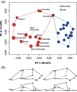
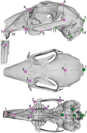
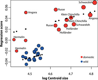
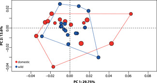
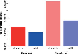
Domestication leads to phenotypic characteristics that have been described to be similar across species. However, this "domestication syndrome" has been subject to debate, related to a lack of evidence for certain characteristics in many species. Here we review diverse literature and provide new data on cranial shape changes due to domestication in the European rabbit (Oryctolagus cuniculus) as a preliminary case study, thus contributing novel evidence to the debate. We quantified cranial shape of 30 wild and domestic rabbits using micro-computed tomography scans and three-dimensional geometric morphometrics. The goal was to test (1) if the domesticates exhibit shorter and broader snouts, smaller teeth, and smaller braincases than their wild counterparts; (2) to what extent allometric scaling is responsible for cranial shape variation; (3) if there is evidence for more variation in the neural crest-derived parts of the cranium compared with those derived of the mesoderm, in accordance with the "neural crest hypothesis." Our own data are consistent with older literature records, suggesting that although there is evidence for some cranial characteristics of the "domestication syndrome" in rabbits, facial length is not reduced. In accordance with the "neural crest hypothesis," we found more shape variation in neural crest versus mesoderm-derived parts of the cranium. Within the domestic group, allometric scaling relationships of the snout, the braincase, and the teeth shed new light on ubiquitous patterns among related taxa. This study-albeit preliminary due to the limited sample size-adds to the growing evidence concerning nonuniform patterns associated with domestication.

Keywords: Oryctolagus cuniculus; allometry; cranium; modularity.


Conflict of interest statement

The authors declare no conflict of interest.

Figures

Figure 1
Figure 1
Results of allometry‐included principal component analysis (PCA) comparing cranial shape in domestic and wild rabbits. In the cranial shape morphospace (a) colors denote the wild (blue) and the domestic (red) groups, and plotted points are scaled to centroid size. Names of domestic breeds (where known) are given. Convex hulls outline the portion of morphospace occupied by the wild and the domestic group, respectively. Wireframes (b) show the skull shapes of the most extreme specimens (in black) relative to the consensus shape (in gray), representing the most negative PC1 scores (left) and most positive PC1 scores (right). Depicted are wireframes in lateral (top) and dorsal (bottom) views (see also Figure 2).
Figure 2
Figure 2
Landmarks and wireframes used in this study. Landmarks are depicted on a surface reconstruction of a wild rabbit (I.f.H. 5188, mirrored) in lateral (top), dorsal (middle), and ventral (bottom) views, and correspond to numbers in Table 1. The gray dashed inlet box depicts landmark No. 1 on the anterior side of the skull. Colors denote the embryonic origin of the bone tissue on which the landmarks are set, following Mishina and Snider (2014): purple, neural crest origin; green, mesoderm origin (see also Table 1). Light shading of points with a dashed outline indicates landmarks that cannot be depicted properly in the respective aspect, but are shown to illustrate the wireframe (black lines connecting the landmarks).
Figure 3
Figure 3
Multivariate regression of cranial shape against log‐transformed centroid size. The regression score represents shape variation attributed to size variation. Colors denote the wild (blue) and the domestic (red) groups, and plotted points are scaled to centroid size. Black dots represent the predicted values of the analysis of covariance, demonstrating the two groups have different allometric trajectories.
Figure 4
Figure 4
Results of allometry‐adjusted principal component analysis (PCA) comparing cranial shape in domestic and wild rabbits. Colors denote the wild (blue) and the domestic (red) groups, and plotted points are scaled to centroid size. The morphological disparity of domesticates is significantly higher than the wild, as demonstrated by the convex hulls outlining the portion of morphospace occupied by the two groups.
Figure 5
Figure 5
Comparison of Procrustes variances of the mesoderm (solid bars) and the neural crest (transparent)‐derived parts of the skull in wild and domestic rabbits (see also Figure 2). Permutation tests showed that the shape of both modules is by trend more variable in the domestic group compared to the wild one, and that the neural crest is significantly different between wild and domestic rabbits.

Similar articles

Cited by

References

    1. Adams, D. C. , Collyer, M. L. , Kaliontzopoulou, A. , & Baken, E. K. 2022. Geomorph: Software for geometric morphometric analyses. R package version 4.0.2. https://cran.r-project.org/package=geomorph
    1. Balcarcel, A. , Geiger, M. , Clauss, M. , & Sánchez‐Villagra, M. (2021). The mammalian brain under domestication: Discovering patterns after a century of old and new analyses. Journal of Experimental Zoology Part B: Molecular and Developmental Evolution , 1–24. - PMC - PubMed
    1. Balcarcel, A. , Sánchez‐Villagra, M. , Segura, V. , & Evin, A. (2021). Singular patterns of skull shape and brain size change in the domestication of South American camelids. Journal of Mammalogy, 102(1), 220–235.
    1. Bauchot, R. (1978). Encephalization in vertebrates. Brain, Behavior and Evolution, 15(1), 1–18. - PubMed
    1. Böhmer, C. , & Böhmer, E. (2017). Shape variation in the craniomandibular system and prevalence of dental problems in domestic rabbits: a case study in Evolutionary Veterinary Science. Veterinary Sciences, 4(1), 5. - PMC - PubMed

Publication types