

Feline intralenticular Encephalitozoon cuniculi: three cases from California
- PMID: 35935143
- PMCID: PMC9350498
- DOI: 10.1177/20551169221106721
Feline intralenticular Encephalitozoon cuniculi: three cases from California
Abstract
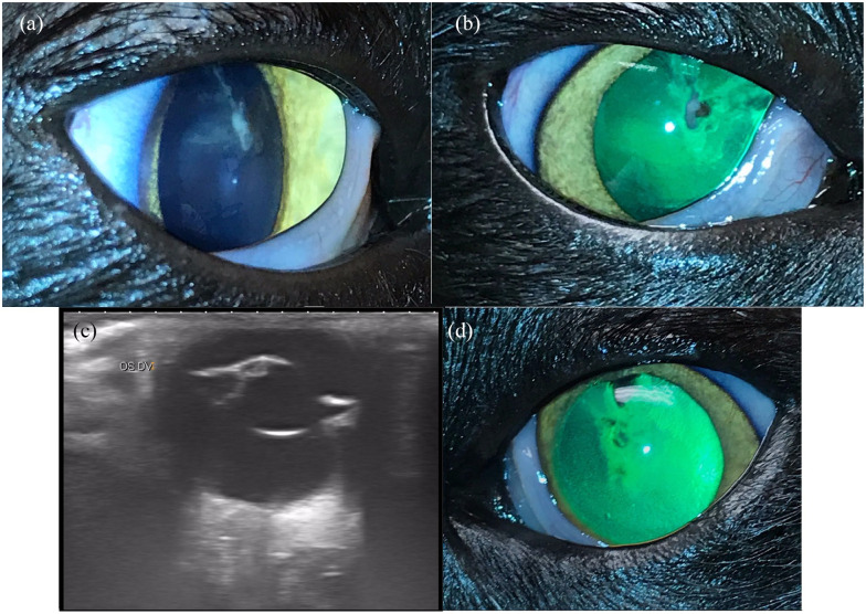
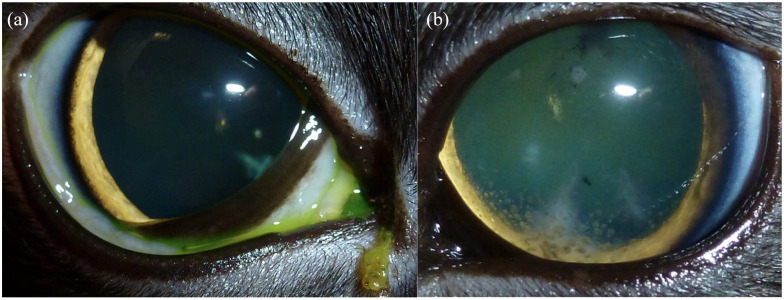
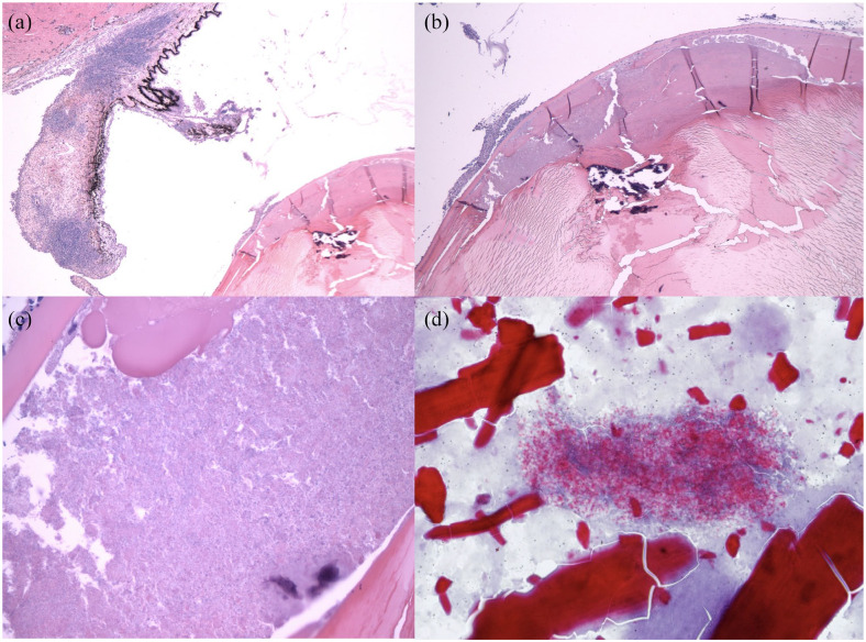
Case series summary: Three domestic shorthair cats from California presented to veterinary ophthalmologists with immature cataracts. Other presenting clinical signs included corneal edema, anisocoria, anterior uveitis, elevated intraocular pressure, blepharospasm and/or lethargy. All patients were immunocompromised due to concurrent diseases and/or immunomodulatory drugs. Diagnostics included serial comprehensive ophthalmic examinations with tonometry, ocular ultrasound, electroretinogram and testing for other causes of feline uveitis. Testing for Encephalitozoon cuniculi included serology, histopathology and/or PCR of aqueous humor, lens material or paraffin-embedded whole eye. Treatments included antiparasitic medication, anti-inflammatory medication and supportive care in all three cases. Surgical treatment included enucleation (one case), bilateral phacoemulsification and unilateral intraocular lens placement (one case) and bilateral phacoemulsification with bilateral endolaser ciliary body ablation and bilateral intraocular lens implantation (one case). Both cats for which serologic testing for E cuniculi was performed were positive (1:64-1:4096). In all cats, diagnosis of intraocular E cuniculi was based on at least one of the following: lens histopathology or PCR of aqueous humor, lens material or paraffin-embedded ocular tissue. The clinical visual outcome was best in the patient undergoing phacoemulsification at the earliest stage of the cataract.
Relevance and novel information: Encephalitozoon cuniculi should be considered as a differential cause of cataracts and uveitis in cats in California, the rest of the USA and likely worldwide.
Keywords: Encephalitozoon cuniculi; cataracts; phacoemulsification; uveitis.
© The Author(s) 2022.
Conflict of interest statement
Conflict of interest: The authors declared no potential conflicts of interest with respect to the research, authorship, and/or publication of this article.
Figures
References
-
- Bohne W, Böttcher K, Groß U. The parasitophorous vacuole of Encephalitozoon cuniculi: biogenesis and characteristics of the host cell-pathogen interface. Int J Med Microbiol 2011; 301: 395–399. - PubMed
-
- Harcourt-Brown F. Infectious diseases of domestic rabbits. In: Harcourt-Brown F. (ed). Textbook of rabbit medicine. St Louis, MO: 2002, pp 361–385.
-
- Jeklova E, Leva L, Kovarcik K, et al.. Experimental oral and ocular Encephalitozoon cuniculi infection in rabbits. Parasitology 2010; 137: 1749–1757. - PubMed
Publication types
Grants and funding
LinkOut - more resources
Full Text Sources
Miscellaneous