

Case Reports
doi: 10.1016/j.jaccas.2022.05.018.
Spontaneous Coronary Artery Dissection in an Orthotopic Heart Transplant Recipient
Affiliations
- PMID: 35935148
- PMCID: PMC9350898
- DOI: 10.1016/j.jaccas.2022.05.018
Item in Clipboard
Case Reports
Spontaneous Coronary Artery Dissection in an Orthotopic Heart Transplant Recipient
JACC Case Rep.
.
Abstract
We present the case of acute myocardial infarction secondary to spontaneous coronary artery dissection in a patient 2 weeks post orthotopic heart transplantation. (Level of Difficulty: Advanced.).
Keywords: FMD, fibromuscular dysplasia; ICH, intracranial hemorrhage; LGE, late gadolinium enhancement; LV, left ventricular; SCAD, spontaneous coronary artery dissection; TTE, transthoracic echocardiogram; acute coronary syndrome; cardiac transplant; dissection; myocardial infarction; systolic heart failure.
© 2022 The Authors.
Conflict of interest statement
The authors have reported that they have no relationships relevant to the contents of this paper to disclose.
Figures

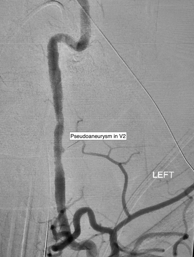
Donor Digital Subtraction Angiography of the Left Vertebral Artery Donor digital subtraction angiography demonstrating irregularities in the vertebral arteries and a small pseudoaneurysm in V2 of the left vertebral artery.
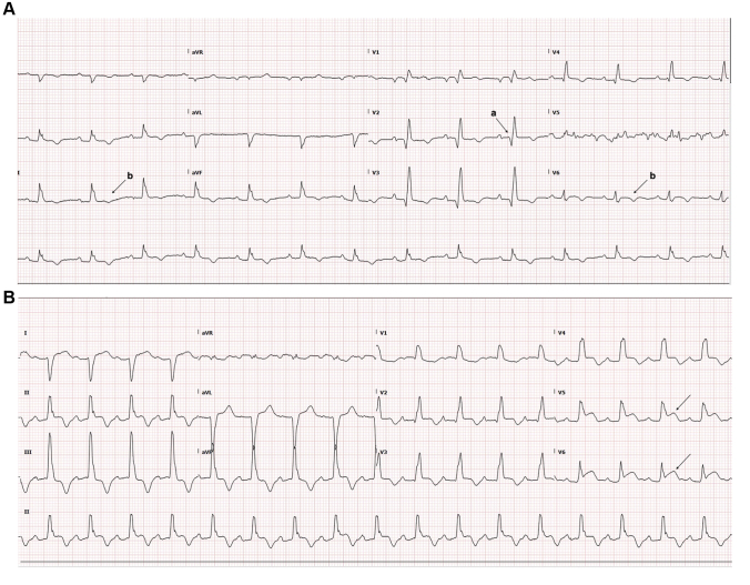
Electrocardiograms (A) Electrocardiogram day 13 post-transplant demonstrating anterior Q waves (a) and diffuse T-wave inversion (b). (B) Electrocardiogram day 8 post-transplant showing ST-segment elevation in V5 and V6(arrows).
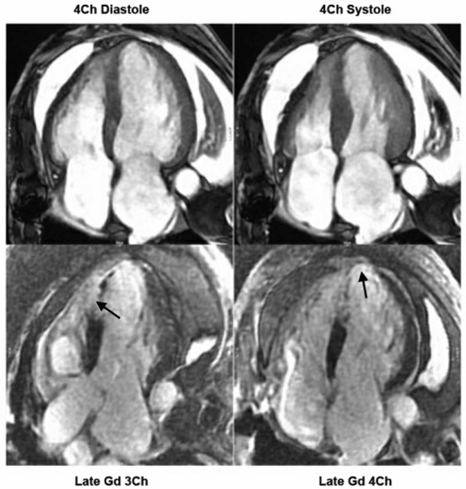
Cardiac Magnetic Resonance Imaging Demonstrating Wall Thinning, Hypokinesis, and Transmural Late Gadolinium Enhancement of the Apical and Anteroseptal Segments of the Left Ventricle Cardiac magnetic resonance imaging demonstrating wall thinning, hypokinesis, and transmural late gadolinium (Gd) enhancement (arrows) of the apical and anteroseptal segments of the left ventricle. Ch = chamber.
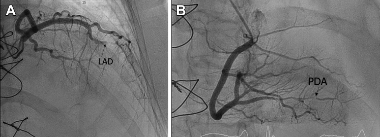
Coronary Angiograms (A) Coronary angiogram demonstrating a sub-totally occluded mid left anterior descending coronary artery (LAD). (B) Coronary angiogram demonstrating spontaneous coronary artery dissection in the distal posterior descending coronary artery (PDA).
References
-
- Saw J., Mancini G.B.J., Humphries K.H. Contemporary review on spontaneous coronary artery dissection. J Am Coll Cardiol. 2016;68:297–312. - PubMed
-
- Theertham A., Niazi K., Moreyra A. Spontaneous coronary artery dissection in heart transplant recipient. J Am Coll Cardiol. 2020;75(11_Supplement_1):2980.
-
- Tsimikas S., Giordano F.J., Tarazi R.Y., et al. Spontaneous coronary artery dissection in patients with renal transplantation. J Invasive Cardiol. 1999;11:316–321. - PubMed
-
- Gornik H.L., Persu A., Adlam D., et al. First international consensus on the diagnosis and management of fibromuscular dysplasia. Vasc Med. 2019;24(2):164–189. - PubMed
Publication types
LinkOut - more resources
Full Text Sources
