Enhancing epitope of PEDV spike protein
- PMID: 35935230
- PMCID: PMC9355140
- DOI: 10.3389/fmicb.2022.933249
Enhancing epitope of PEDV spike protein
Abstract
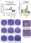
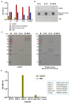
Porcine epidemic diarrhea virus (PEDV) is the causative agent of a highly contagious enteric disease of pigs characterized by diarrhea, vomiting, and severe dehydration. PEDV infects pigs of all ages, but neonatal pigs during the first week of life are highly susceptible; the mortality rates among newborn piglets may reach 80-100%. Thus, PEDV is regarded as one of the most devastating pig viruses that cause huge economic damage to pig industries worldwide. Vaccination of sows and gilts at the pre-fertilization or pre-farrowing stage is a good strategy for the protection of suckling piglets against PEDV through the acquisition of the lactating immunity. However, vaccination of the mother pigs for inducing a high level of virus-neutralizing antibodies is complicated with unstandardized immunization protocol and unreliable outcomes. Besides, the vaccine may also induce enhancing antibodies that promote virus entry and replication, so-called antibody-dependent enhancement (ADE), which aggravates the disease upon new virus exposure. Recognition of the virus epitope that induces the production of the enhancing antibodies is an existential necessity for safe and effective PEDV vaccine design. In this study, the enhancing epitope of the PEDV spike (S) protein was revealed for the first time, by using phage display technology and mouse monoclonal antibody (mAbG3) that bound to the PEDV S1 subunit of the S protein and enhanced PEDV entry into permissive Vero cells that lack Fc receptor. The phages displaying mAbG3-bound peptides derived from the phage library by panning with the mAbG3 matched with several regions in the S1-0 sub-domain of the PEDV S1 subunit, indicating that the epitope is discontinuous (conformational). The mAbG3-bound phage sequence also matched with a linear sequence of the S1-BCD sub-domains. Immunological assays verified the phage mimotope results. Although the molecular mechanism of ADE caused by the mAbG3 via binding to the newly identified S1 enhancing epitope awaits investigation, the data obtained from this study are helpful and useful in designing a safe and effective PEDV protein subunit/DNA vaccine devoid of the enhancing epitope.
Keywords: antibody-dependent enhancement; enhancing epitope; phage display technology; phage mimotope; phage panning; porcine epidemic diarrhea; porcine epidemic diarrhea virus; spike protein.
Copyright © 2022 Thavorasak, Chulanetra, Glab-ampai, Mahasongkram, Sae-lim, Teeranitayatarn, Songserm, Yodsheewan, Nilubol, Chaicumpa and Sookrung.
Conflict of interest statement
KT was employed by the MORENA Solution Company. The remaining authors declare that the research was conducted in the absence of any commercial or financial relationships that could be construed as a potential conflict of interest.
Figures


Similar articles
-
Novel Neutralizing Epitope of PEDV S1 Protein Identified by IgM Monoclonal Antibody.Viruses. 2022 Jan 11;14(1):125. doi: 10.3390/v14010125. Viruses. 2022. PMID: 35062329 Free PMC article.
-
Cell Attachment Domains of the Porcine Epidemic Diarrhea Virus Spike Protein Are Key Targets of Neutralizing Antibodies.J Virol. 2017 May 26;91(12):e00273-17. doi: 10.1128/JVI.00273-17. Print 2017 Jun 15. J Virol. 2017. PMID: 28381581 Free PMC article.
-
S1 domain of the porcine epidemic diarrhea virus spike protein as a vaccine antigen.Virol J. 2016 Apr 1;13:57. doi: 10.1186/s12985-016-0512-8. Virol J. 2016. PMID: 27036203 Free PMC article.
-
Porcine epidemic diarrhea virus (PEDV): An update on etiology, transmission, pathogenesis, and prevention and control.Virus Res. 2020 Sep;286:198045. doi: 10.1016/j.virusres.2020.198045. Epub 2020 Jun 2. Virus Res. 2020. PMID: 32502552 Free PMC article. Review.
-
Host Factors Affecting Generation of Immunity Against Porcine Epidemic Diarrhea Virus in Pregnant and Lactating Swine and Passive Protection of Neonates.Pathogens. 2020 Feb 18;9(2):130. doi: 10.3390/pathogens9020130. Pathogens. 2020. PMID: 32085410 Free PMC article. Review.
Cited by
-
The Characterization and Pathogenicity of a Recombinant Porcine Epidemic Diarrhea Virus Variant ECQ1.Viruses. 2023 Jun 30;15(7):1492. doi: 10.3390/v15071492. Viruses. 2023. PMID: 37515178 Free PMC article.
-
Research progress of porcine epidemic diarrhea virus S protein.Front Microbiol. 2024 May 30;15:1396894. doi: 10.3389/fmicb.2024.1396894. eCollection 2024. Front Microbiol. 2024. PMID: 38873162 Free PMC article. Review.
-
Midnolin inhibits coronavirus proliferation by degrading viral proteins.J Virol. 2025 Jun 17;99(6):e0036625. doi: 10.1128/jvi.00366-25. Epub 2025 May 29. J Virol. 2025. PMID: 40439407 Free PMC article.
-
Identification of host proteins interacting with the E protein of porcine epidemic diarrhea virus.Front Microbiol. 2024 Mar 21;15:1380578. doi: 10.3389/fmicb.2024.1380578. eCollection 2024. Front Microbiol. 2024. PMID: 38577683 Free PMC article.
-
Surface display of the COE antigen of porcine epidemic diarrhoea virus on Bacillus subtilis spores.Microb Biotechnol. 2024 Jul;17(7):e14518. doi: 10.1111/1751-7915.14518. Microb Biotechnol. 2024. PMID: 38953907 Free PMC article.
References
-
- Chang C. Y., Cheng I. C., Chang Y. C., Tsai P. S., Lai S. Y., Huang Y. L., et al. . (2019). Identification of neutralizing monoclonal antibodies targeting novel conformational epitopes of the porcine epidemic diarrhoea virus spike protein. Sci. Rep. 9, 2529. doi: 10.1038/s41598-019-39844-5, PMID: - DOI - PMC - PubMed
-
- Chang C. Y., Wang Y.-S., Wu J.-F., Yang T.-J., Chang Y.-C., Chae C., et al. . (2021). Generation and characterization of a spike glycoprotein domain A-specific neutralizing single-chain variable fragment against porcine epidemic diarrhea virus. Vaccines. 9, 833. doi: 10.3390/vaccines9080833, PMID: - DOI - PMC - PubMed
-
- Chang S. H., Bae J. L., Kang T. J., Kim J., Chung G. H., Lim C. W., et al. . (2002). Identification of the epitope region capable of inducing neutralizing antibodies against the porcine epidemic diarrhea virus. Mol. Cells 14, 295–299. - PubMed
-
- Chareonsirisuthigul T., Kalayanarooj S., Ubol S. (2007). Dengue virus (DENV) antibody-dependent enhancement of infection upregulates the production of anti-inflammatory cytokines but suppresses anti-DENV free radical and pro-inflammatory cytokine production, in THP-1 cells. J. Gen. Virol. 88, 365–375. doi: 10.1099/vir.0.82537-0, PMID: - DOI - PubMed
LinkOut - more resources
Full Text Sources
Molecular Biology Databases

