

SARS-CoV-2 Nucleocapsid Protein Has DNA-Melting and Strand-Annealing Activities With Different Properties From SARS-CoV-2 Nsp13
- PMID: 35935242
- PMCID: PMC9354549
- DOI: 10.3389/fmicb.2022.851202
SARS-CoV-2 Nucleocapsid Protein Has DNA-Melting and Strand-Annealing Activities With Different Properties From SARS-CoV-2 Nsp13
Abstract
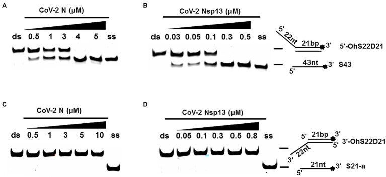
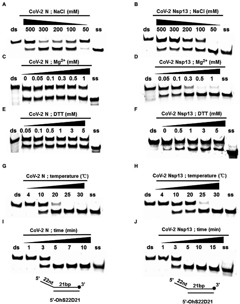
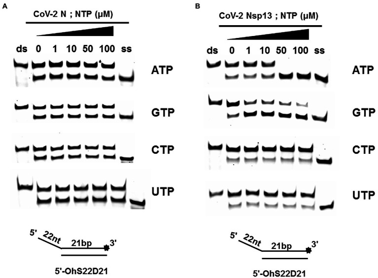
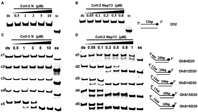
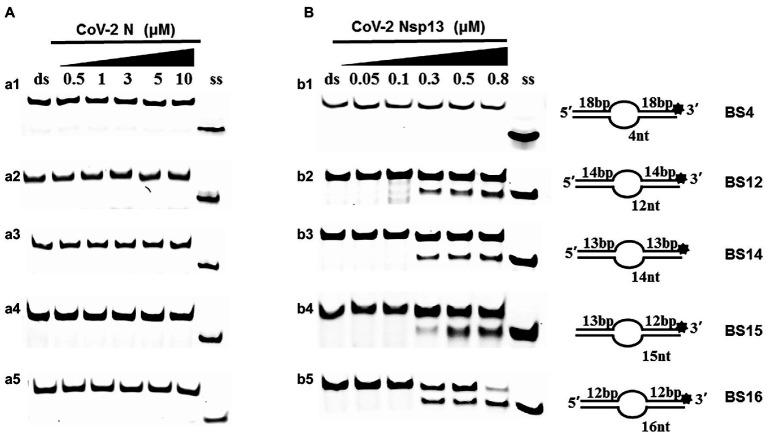
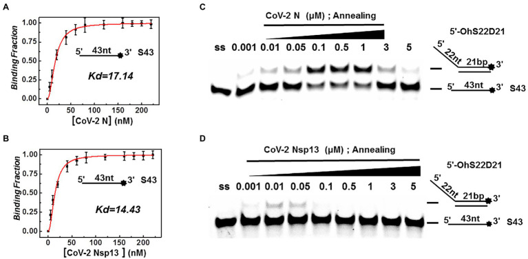
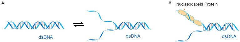
Since December 2019, severe acute respiratory syndrome coronavirus 2 (SARS-CoV-2) has spread throughout the world and has had a devastating impact on health and economy. The biochemical characterization of SARS-CoV-2 proteins is important for drug design and development. In this study, we discovered that the SARS-CoV-2 nucleocapsid protein can melt double-stranded DNA (dsDNA) in the 5'-3' direction, similar to SARS-CoV-2 nonstructural protein 13. However, the unwinding activity of SARS-CoV-2 nucleocapsid protein was found to be more than 22 times weaker than that of SARS-CoV-2 nonstructural protein 13, and the melting process was independent of nucleoside triphosphates and Mg2+. Interestingly, at low concentrations, the SARS-CoV-2 nucleocapsid protein exhibited a stronger annealing activity than SARS-CoV-2 nonstructural protein 13; however, at high concentrations, it promoted the melting of dsDNA. These findings have deepened our understanding of the SARS-CoV-2 nucleocapsid protein and will help provide novel insights into antiviral drug development.
Keywords: Nsp13; SARS-CoV-2; helicase; nucleocapsid protein; unwinding.
Copyright © 2022 Zhang, Xie, Lan, Li, Tian, Zhang, Tian, Yang, Zhou, Qiu, Lu and Liu.
Conflict of interest statement
The authors declare that the research was conducted in the absence of any commercial or financial relationships that could be construed as a potential conflict of interest.
Figures
Similar articles
-
Multiple enzymatic activities associated with severe acute respiratory syndrome coronavirus helicase.J Virol. 2004 Jun;78(11):5619-32. doi: 10.1128/JVI.78.11.5619-5632.2004. J Virol. 2004. PMID: 15140959 Free PMC article.
-
Biochemical Characterization of Middle East Respiratory Syndrome Coronavirus Helicase.mSphere. 2016 Sep 7;1(5):e00235-16. doi: 10.1128/mSphere.00235-16. eCollection 2016 Sep-Oct. mSphere. 2016. PMID: 27631026 Free PMC article.
-
Biochemical analysis of SARS-CoV-2 Nsp13 helicase implicated in COVID-19 and factors that regulate its catalytic functions.J Biol Chem. 2023 Mar;299(3):102980. doi: 10.1016/j.jbc.2023.102980. Epub 2023 Feb 4. J Biol Chem. 2023. PMID: 36739951 Free PMC article.
-
Overview of SARS-CoV-2 genome-encoded proteins.Sci China Life Sci. 2022 Feb;65(2):280-294. doi: 10.1007/s11427-021-1964-4. Epub 2021 Aug 10. Sci China Life Sci. 2022. PMID: 34387838 Free PMC article. Review.
-
The SARS-CoV-2 Nucleocapsid Protein and Its Role in Viral Structure, Biological Functions, and a Potential Target for Drug or Vaccine Mitigation.Viruses. 2021 Jun 10;13(6):1115. doi: 10.3390/v13061115. Viruses. 2021. PMID: 34200602 Free PMC article. Review.
Cited by
-
Structural domains of SARS-CoV-2 nucleocapsid protein coordinate to compact long nucleic acid substrates.Nucleic Acids Res. 2023 Jan 11;51(1):290-303. doi: 10.1093/nar/gkac1179. Nucleic Acids Res. 2023. PMID: 36533523 Free PMC article.
-
Structural basis for the participation of the SARS-CoV-2 nucleocapsid protein in the template switch mechanism and genomic RNA reorganization.J Biol Chem. 2024 Nov;300(11):107834. doi: 10.1016/j.jbc.2024.107834. Epub 2024 Sep 27. J Biol Chem. 2024. PMID: 39343000 Free PMC article. Review.
-
Affinity Tag-Free Purification of SARS-CoV-2 N Protein and Its Crystal Structure in Complex with ssDNA.Biomolecules. 2024 Nov 30;14(12):1538. doi: 10.3390/biom14121538. Biomolecules. 2024. PMID: 39766245 Free PMC article.
References
-
- Adedeji A. O., Singh K., Calcaterra N. E., DeDiego M. L., Enjuanes L., Weiss S., et al. . (2012). Severe acute respiratory syndrome coronavirus replication inhibitor that interferes with the nucleic acid unwinding of the viral helicase. Antimicrob. Agents Chemother. 56, 4718–4728. doi: 10.1128/AAC.00957-12, PMID: - DOI - PMC - PubMed
-
- Carlson C. R., Asfaha J. B., Ghent C. M., Howard C. J., Hartooni N., Safari M., et al. . (2020). Phosphoregulation of phase separation by the SARS-CoV-2 N protein suggests a biophysical basis for its dual functions. Mol. Cell 80, 1092–1103. doi: 10.1016/j.molcel.2020.11.025, PMID: - DOI - PMC - PubMed
LinkOut - more resources
Full Text Sources
Miscellaneous