

Diagnostic Value of Image Features of Magnetic Resonance Imaging in Intracranial Hemorrhage and Cerebral Infarction
- PMID: 35935302
- PMCID: PMC9296345
- DOI: 10.1155/2022/6495568
Diagnostic Value of Image Features of Magnetic Resonance Imaging in Intracranial Hemorrhage and Cerebral Infarction
Abstract
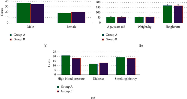
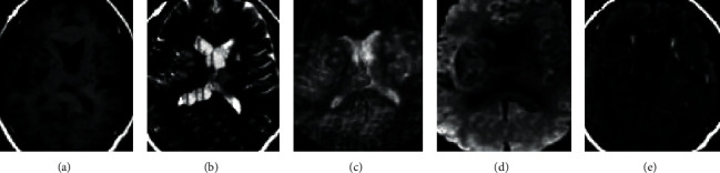
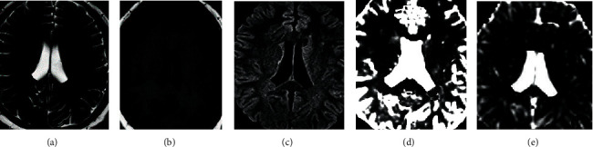
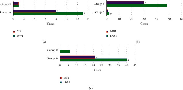
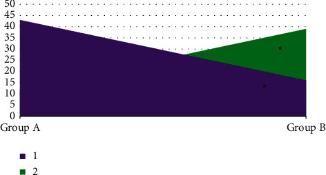
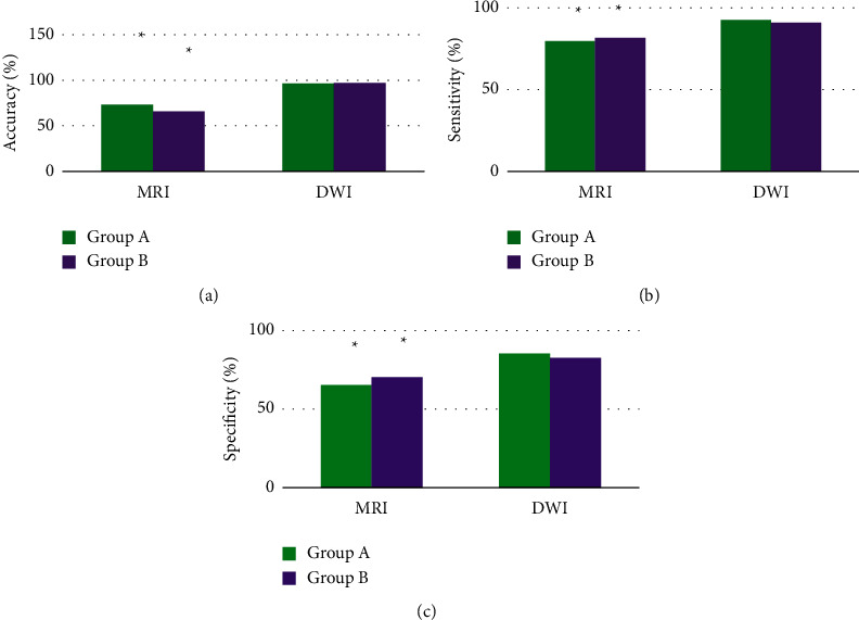
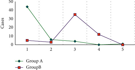
This study aimed to investigate the differential diagnosis value of routine magnetic resonance imaging (MRI) and magnetic resonance diffusion-weighted imaging (DWI) in hyperacute intracranial hemorrhage (HICH) and hyperacute cerebral infarction (HCI). Fifty-five patients with HICH were set as group A, and 55 patients with HCI were selected as group B. All the patients underwent routine MRI and DWI examinations. The morphological distribution and signal characteristics (low, high, or mixed) of the lesions in the two groups were recorded. The diagnostic accuracy, sensitivity, and specificity of routine MRI and DWI were compared for distinguishing HICH and HCI. The results suggested that the lesions in patients with HICH were mainly manifested as mixed signals (40 cases), while those in patients with HCI showed high signals (48 cases). HICH occurred in the basal ganglia in 44 cases, in the brain stem in 6 cases, in the cerebellum in 4 cases, in the cerebral cortex in 0 cases, and in the corpus callosum in 1 case. HCI occurred in the basal ganglia area, brain stem, cerebellum, cerebral cortex, and corpus callosum in 5, 3, 35, 12, and 0 cases, respectively. The diagnostic accuracy, specificity, and sensitivity of DWI for HICH and HCI were significantly higher than those of routine MRI (P < 0.05). It was indicated that compared with routine MRI, DWI was more effective in the diagnosis of HICH and HCI, with clearer and more accurate images and better diagnostic performance.
Copyright © 2022 Wencai Tang et al.
Conflict of interest statement
The authors declare that they have no conflicts of interest.
Figures
Similar articles
-
[Early diagnostic significance and dynamic pattern of DWI compared with conventional MRI in newborns with neonatal cerebral infarction].Zhonghua Er Ke Za Zhi. 2007 May;45(5):360-4. Zhonghua Er Ke Za Zhi. 2007. PMID: 17697623 Chinese.
-
[Application of diffusion-weighted and perfusion magnetic resonance imaging in definition of the ischemic penumbra in hyperacute cerebral infarction].Zhonghua Yi Xue Za Zhi. 2003 Jun 10;83(11):952-7. Zhonghua Yi Xue Za Zhi. 2003. PMID: 12899795 Chinese.
-
[Early assessment of severe hypoxic-ischemic encephalopathy in neonates by diffusion-weighted magnetic resonance imaging techniques and its significance].Zhonghua Er Ke Za Zhi. 2007 Nov;45(11):843-7. Zhonghua Er Ke Za Zhi. 2007. PMID: 18282417 Chinese.
-
18F-FDG PET/CT and whole-body MRI diagnostic performance in M staging for non-small cell lung cancer: a systematic review and meta-analysis.Eur Radiol. 2020 Jul;30(7):3641-3649. doi: 10.1007/s00330-020-06703-1. Epub 2020 Mar 3. Eur Radiol. 2020. PMID: 32125513
-
Application and progress of highcontent imaging in molecular biology.Biotechnol J. 2023 Dec;18(12):e2300170. doi: 10.1002/biot.202300170. Epub 2023 Sep 11. Biotechnol J. 2023. PMID: 37639283 Review.
References
MeSH terms
LinkOut - more resources
Full Text Sources
Medical
Research Materials
