

The Value of Dynamic Contrast-Enhanced Magnetic Resonance Imaging (DCE-MRI) in the Differentiation of Pseudoprogression and Recurrence of Intracranial Gliomas
- PMID: 35935318
- PMCID: PMC9337951
- DOI: 10.1155/2022/5680522
The Value of Dynamic Contrast-Enhanced Magnetic Resonance Imaging (DCE-MRI) in the Differentiation of Pseudoprogression and Recurrence of Intracranial Gliomas
Abstract
Objective: The objective of this study was to determine the value of dynamic contrast-enhanced magnetic resonance imaging (DCE-MRI) in assessing postoperative changes in intracranial gliomas.
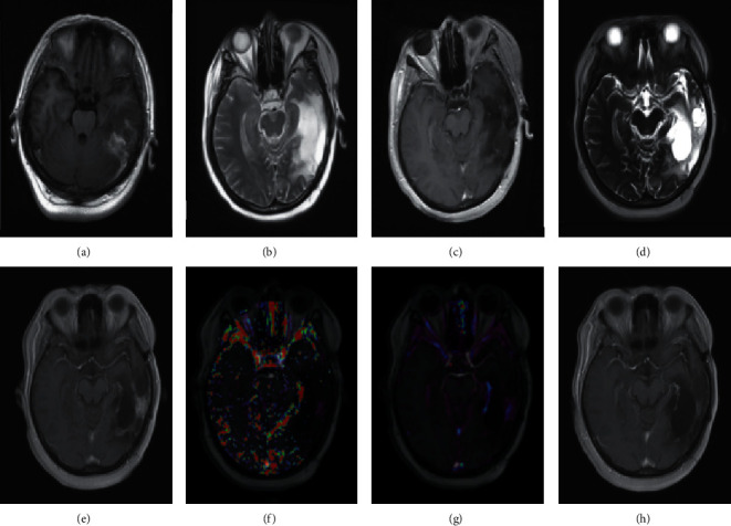
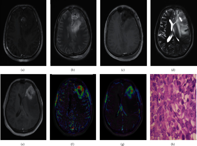
Method: A total of fifty-one patients who had new enhanced lesions after surgical resection followed by standard radiotherapy and chemotherapy were collected retrospectively from October 2014 to June 2021. The patients were divided into a pseudoprogression group (15 cases) and a recurrence group (36 cases) according to the pathological results of the second operation or a follow-up of more than six months. The follow-up data of all patients were complete, and DCE-MRI was performed. The images were processed to obtain the quantitative parameters K trans, Ve, and Kep and the semiquantitative parameter iAUC, which were analysed with relevant statistical software.
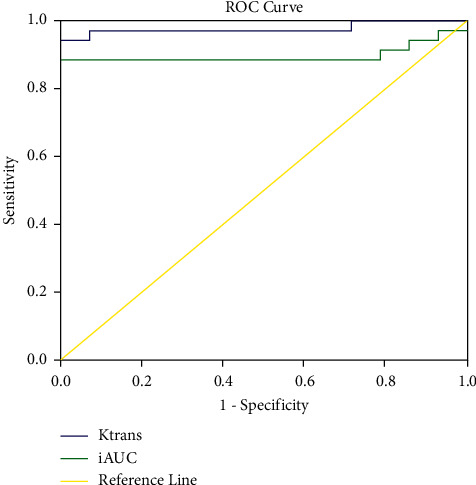
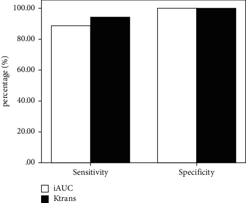
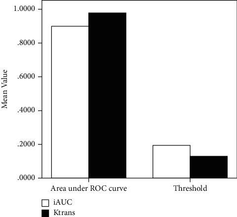
Results: First, the difference in K trans and iAUC values between the two groups was statistically significant (P < 0.05), and the difference in Ve and Kep values was not statistically significant (P > 0.05). Second, by comparing the area under the curve, threshold, sensitivity and specificity of K trans, and iAUC, it was found that the iAUC threshold value was slightly higher than that of K trans, and the specificity of K trans was equal to that of iAUC, while the area under the curve and sensitivity of K trans were higher than those of iAUC. Third, K trans and iAUC had high accuracy in diagnosing recurrence and pseudoprogression, and K trans had higher accuracy than iAUC.
Conclusion: In this study, DCE-MRI has a certain diagnostic value in the early differentiation of recurrence and pseudoprogression, offering a new method for the diagnosis and assessment of gliomas after surgery.
Copyright © 2022 Hui Jing et al.
Conflict of interest statement
The authors declare that they have no conflicts of interests.
Figures
Similar articles
-
[The value of DCE-MRI in predicting IDH gene mutation of high-grade gliomas].Zhonghua Yi Xue Za Zhi. 2019 Oct 22;99(39):3105-3109. doi: 10.3760/cma.j.issn.0376-2491.2019.39.013. Zhonghua Yi Xue Za Zhi. 2019. PMID: 31648456 Chinese.
-
Distinguishing recurrent high-grade gliomas from radiation injury: a pilot study using dynamic contrast-enhanced MR imaging.Acad Radiol. 2011 May;18(5):575-83. doi: 10.1016/j.acra.2011.01.018. Epub 2011 Mar 21. Acad Radiol. 2011. PMID: 21419671 Clinical Trial.
-
Diagnostic Values of DCE-MRI and DSC-MRI for Differentiation Between High-grade and Low-grade Gliomas: A Comprehensive Meta-analysis.Acad Radiol. 2018 Mar;25(3):338-348. doi: 10.1016/j.acra.2017.10.001. Epub 2017 Dec 6. Acad Radiol. 2018. PMID: 29223713
-
Glioma grading capability: comparisons among parameters from dynamic contrast-enhanced MRI and ADC value on DWI.Korean J Radiol. 2013 May-Jun;14(3):487-92. doi: 10.3348/kjr.2013.14.3.487. Epub 2013 May 2. Korean J Radiol. 2013. PMID: 23690718 Free PMC article.
-
Diagnostic accuracy of dynamic contrast-enhanced perfusion MRI in stratifying gliomas: A systematic review and meta-analysis.Cancer Med. 2019 Sep;8(12):5564-5573. doi: 10.1002/cam4.2369. Epub 2019 Aug 7. Cancer Med. 2019. PMID: 31389669 Free PMC article.
Cited by
-
Review of tracer kinetic models in evaluation of gliomas using dynamic contrast-enhanced imaging.Front Oncol. 2024 Jun 14;14:1380793. doi: 10.3389/fonc.2024.1380793. eCollection 2024. Front Oncol. 2024. PMID: 38947892 Free PMC article. Review.
References
Publication types
MeSH terms
Substances
LinkOut - more resources
Full Text Sources
