

Small vessel vasculitis associated with culture-negative infective endocarditis related to a cardiac device: a case report
- PMID: 35935394
- PMCID: PMC9351725
- DOI: 10.1093/ehjcr/ytac294
Small vessel vasculitis associated with culture-negative infective endocarditis related to a cardiac device: a case report
Abstract
Background: Culture-negative endocarditis is uncommon, occurring in less than a third of all cases of infective endocarditis (IE). Culture-negative IE related to a cardiac device is an even greater diagnostic challenge due to its insidious presentation, with onset of symptoms ranging between 3 and 12 months after device implantation. Sensitivity of the modified Duke's criteria remains low in culture-negative and cardiac device-related IE (CDRIE) since classical signs and symptoms of IE are often absent. Small vessel vasculitis has been reported as an immune response to IE. Recognizing immunological phenomenon related to IE is of paramount clinical importance, prompting the search for an underlying infection and avoiding the use of immunosuppressive medications which would otherwise result in an adverse outcome.
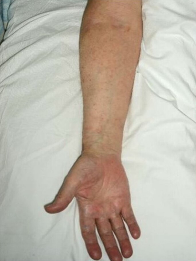
Case summary: An 81-year-old Caucasian male presented to the ambulatory medical unit with a two-week history of a symmetrical, generalized purpuric rash. He had an indwelling permanent pacemaker following a transcatheter aortic valve implantation for severe aortic stenosis five years ago. Blood tests showed an iron deficiency anaemia, thrombocytopenia and normal renal function, both CRP and ESR were raised at 61 and 30 mm/hr, respectively. Skin biopsy demonstrated small vessel cutaneous vasculitis. Transthoracic echocardiography revealed a mobile mass measuring 0.9 × 1.7 cm, confirmed on transoesophageal echocardiogram as pacing lead endocarditis. Blood cultures were persistently negative. The patient underwent pacemaker lead extraction, following which the vasculitic rash improved.
Discussion: Blood cultures in IE are more likely to be negative if there is a prior antibiotic administration or causative micro-organisms with limited proliferation which fail to grow in conventional media conditions. Transesophageal echocardiography (TOE) offers improved sensitivity and diagnostic yield when compared to transthoracic echocardiography (TTE) in patients with a high clinical suspicion of CDRIE. The evidence in the literature describing culture-negative IE associated with small vessel vasculitis is limited. However, it is recognized that cutaneous small vessel vasculitis may be associated with an underlying bacterial infection. IE produces an inflammatory response, resulting in the deposition of circulating immune complexes and cutaneous signs which are included in the modified Duke's criteria to aid diagnosis. Management of CDRIE requires a multi-disciplinary team approach with an 'Endocarditis Team.' Pacemaker lead infection requires transvenous lead extraction if it is a newly implanted lead. Locking stylets, extraction sheaths or snare retrieval are usually required in cases of older implanted leads. Surgical lead extraction remains the gold standard for larger vegetations (>20 mm) or associated valve endocarditis.
Keywords: Case report; Cutaneous small vessel vasculitis; Infective endocarditis; Permanent pacemaker; Transesophageal echocardiography; Transthoracic echocardiography.
© The Author(s) 2022. Published by Oxford University Press on behalf of the European Society of Cardiology.
Figures


Similar articles
-
Transcatheter aortic valve replacement associated infective endocarditis case series: broadening the criteria for diagnosis is the need of the hour.BMC Cardiovasc Disord. 2021 Nov 20;21(1):559. doi: 10.1186/s12872-021-02364-0. BMC Cardiovasc Disord. 2021. PMID: 34800994 Free PMC article.
-
Can transthoracic echocardiography be used to a greater extent in the diagnostics of infective endocarditis to avoid unnecessary transoesophageal examinations without jeopardising accuracy?Cardiovasc Ultrasound. 2023 Jan 31;21(1):3. doi: 10.1186/s12947-023-00301-z. Cardiovasc Ultrasound. 2023. PMID: 36717895 Free PMC article.
-
Cardiac device-related infective endocarditis need for lead extraction whatever the device according to the ESC EORP EURO-ENDO registry.Eur Heart J Open. 2023 Jul 3;3(4):oead064. doi: 10.1093/ehjopen/oead064. eCollection 2023 Jul. Eur Heart J Open. 2023. PMID: 37465258 Free PMC article.
-
Infective Endocarditis among Pediatric Patients with Prosthetic Valves and Cardiac Devices: A Review and Update of Recent Emerging Diagnostic and Management Strategies.J Clin Med. 2023 Jul 27;12(15):4941. doi: 10.3390/jcm12154941. J Clin Med. 2023. PMID: 37568344 Free PMC article. Review.
-
A narrative review of diagnosis of infective endocarditis-imaging methods and comparison.Ann Transl Med. 2020 Dec;8(23):1621. doi: 10.21037/atm-20-4555. Ann Transl Med. 2020. PMID: 33437820 Free PMC article. Review.
Cited by
-
Case report: Novel three-dimensional echocardiographic methods mapping aortic root pseudoaneurysm secondary to blood culture-negative endocarditis with bicuspid aortic valve involvement.Front Cardiovasc Med. 2023 Mar 16;10:1138390. doi: 10.3389/fcvm.2023.1138390. eCollection 2023. Front Cardiovasc Med. 2023. PMID: 37008335 Free PMC article.
References
-
- Habib G, Lancellotti P, Antunes MJ, Bongiorni MG, Casalta JP, Del Zotti F, Dulgheru R, El Khoury G, Erba PA, Iung B, Miro JM, Mulder BJ, Plonska-Gosciniak E, Price S, Roos-Hesselink J, Snygg-Martin U, Thuny F, Tornos Mas P, Vilacosta I, Zamorano JL; ESC Scientific Document Group . 2015 ESC guidelines for the management of infective endocarditis. Eur Heart J 2015;36:3075–3128. - PubMed
Publication types
LinkOut - more resources
Full Text Sources
Research Materials
Miscellaneous