

Exosome mediated biological functions within skeletal microenvironment
- PMID: 35935491
- PMCID: PMC9355125
- DOI: 10.3389/fbioe.2022.953916
Exosome mediated biological functions within skeletal microenvironment
Abstract
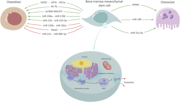
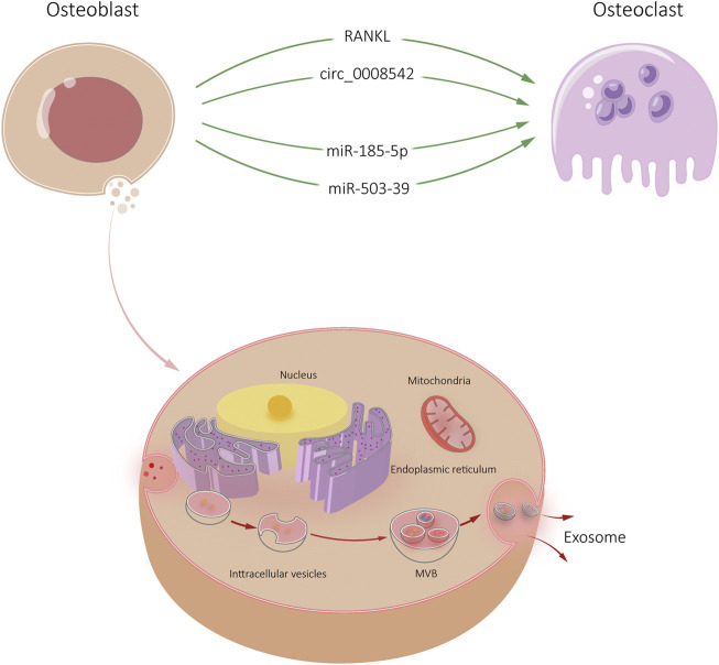
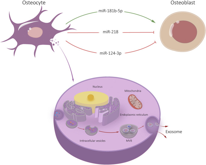
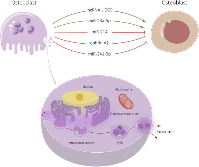
Exosomes are membranous lipid vesicles fused with intracellular multicellular bodies that are released into the extracellular environment. They contain bioactive substances, including proteins, RNAs, lipids, and cytokine receptors. Exosomes in the skeletal microenvironment are derived from a variety of cells such as bone marrow mesenchymal stem cells (BMSCs), osteoblasts, osteoclasts, and osteocytes. Their biological function is key in paracrine or endocrine signaling. Exosomes play a role in bone remodeling by regulating cell proliferation and differentiation. Genetic engineering technology combined with exosome-based drug delivery can therapy bone metabolic diseases. In this review, we summarized the pathways of exosomes derived from different skeletal cells (i.e., BMSCs, osteoblasts, osteocytes, and osteoclasts) regulate the skeletal microenvironment through proteins, mRNAs, and non-coding RNAs. By exploring the role of exosomes in the skeletal microenvironment, we provide a theoretical basis for the clinical treatment of bone-related metabolic diseases, which may lay the foundation to improve bone tumor microenvironments, alleviate drug resistance in patients.
Keywords: bone marrow mesenchymal stem cell; osteoblast; osteoclast; osteocyte; skeletal microenvironment; skeletal related exosomes.
Copyright © 2022 Wang, Zhao, Gao and Zhang.
Conflict of interest statement
The authors declare that the research was conducted in the absence of any commercial or financial relationships that could be construed as a potential conflict of interest.
Figures
References
-
- Aghebati-Maleki L., Dolati S., Zandi R., Fotouhi A., Ahmadi M., Aghebati A., et al. (2019). Prospect of mesenchymal stem cells in therapy of osteoporosis: A review. J. Cell. Physiol. 234 (6), 8570–8578. 10.1002/jcp.27833 PubMed Abstract | 10.1002/jcp.27833 | Google Scholar - DOI - PubMed
-
- Alimirzaie S., Bagherzadeh M., Akbari M. R. (2019). Liquid biopsy in breast cancer: A comprehensive review. Clin. Genet. 95 (6), 643–660. 10.1111/cge.13514 PubMed Abstract | 10.1111/cge.13514 | Google Scholar - DOI - PubMed
-
- Ashwal-Fluss R., Meyer M., Pamudurti N. R., Ivanov A., Bartok O., Hanan M., et al. (2014). circRNA biogenesis competes with pre-mRNA splicing. Mol. Cell 56 (1), 55–66. 10.1016/j.molcel.2014.08.019 PubMed Abstract | 10.1016/j.molcel.2014.08.019 | Google Scholar - DOI - PubMed
-
- Bhushan R., Grunhagen J., Becker J., Robinson P. N., Ott C. E., Knaus P. (2013). miR-181a promotes osteoblastic differentiation through repression of TGF-β signaling molecules. Int. J. Biochem. Cell Biol. 45 (3), 696–705. 10.1016/j.biocel.2012.12.008 PubMed Abstract | 10.1016/j.biocel.2012.12.008 | Google Scholar - DOI - PubMed
-
- Birmingham E., Niebur G. L., McHugh P. E., Shaw G., Barry F. P., McNamara L. M. (2012). Osteogenic differentiation of mesenchymal stem cells is regulated by osteocyte and osteoblast cells in a simplified bone niche. Eur. Cell. Mat. 23, 13–27. 10.22203/ecm.v023a02 PubMed Abstract | 10.22203/ecm.v023a02 | Google Scholar - DOI - PubMed
Publication types
LinkOut - more resources
Full Text Sources