. 2022 Jul 22:10:906295.
doi: 10.3389/fbioe.2022.906295. eCollection 2022.

Transfer mechanism of cell-free synthesized membrane proteins into mammalian cells

Affiliations

Transfer mechanism of cell-free synthesized membrane proteins into mammalian cells

Simon Umbach et al. Front Bioeng Biotechnol. .

Abstract

Nanodiscs are emerging to serve as transfer vectors for the insertion of recombinant membrane proteins into membranes of living cells. In combination with cell-free expression technologies, this novel process opens new perspectives to analyze the effects of even problematic targets such as toxic, hard-to-express, or artificially modified membrane proteins in complex cellular environments of different cell lines. Furthermore, transferred cells must not be genetically engineered and primary cell lines or cancer cells could be implemented as well. We have systematically analyzed the basic parameters of the nanotransfer approach and compared the transfer efficiencies from nanodiscs with that from Salipro particles. The transfer of five membrane proteins was analyzed: the prokaryotic proton pump proteorhodopsin, the human class A family G-protein coupled receptors for endothelin type B, prostacyclin, free fatty acids type 2, and the orphan GPRC5B receptor as a class C family member. The membrane proteins were cell-free synthesized with a detergent-free strategy by their cotranslational insertion into preformed nanoparticles containing defined lipid environments. The purified membrane protein/nanoparticles were then incubated with mammalian cells. We demonstrate that nanodiscs disassemble and only lipids and membrane proteins, not the scaffold protein, are transferred into cell membranes. The process is detectable within minutes, independent of the nanoparticle lipid composition, and the transfer efficiency directly correlates with the membrane protein concentration in the transfer mixture and with the incubation time. Transferred membrane proteins insert in both orientations, N-terminus in and N-terminus out, in the cell membrane, and the ratio can be modulated by engineering. The viability of cells is not notably affected by the transfer procedure, and transferred membrane proteins stay detectable in the cell membrane for up to 3 days. Transferred G-protein coupled receptors retained their functionality in the cell environment as shown by ligand binding, induction of internalization, and specific protein interactions. In comparison to transfection, the cellular membrane protein concentration is better controllable and more uniformly distributed within the analyzed cell population. A further notable difference to transfection is the accumulation of transferred membrane proteins in clusters, presumably determined by microdomain structures in the cell membranes.

Keywords: G-protein coupled receptors; GPCR function; HEK 293 cell; Salipro nanoparticles; cell-free expression; nanodiscs; protein transfer; transfection.


Conflict of interest statement

The authors declare that the research was conducted in the absence of any commercial or financial relationships that could be construed as a potential conflict of interest.

Figures

FIGURE 1
FIGURE 1
SEC profiling of MP/ND samples used for nanotransfer into mammalian cells. The receptors were cotranslationally inserted into preformed NDs, Strep-purified and analyzed with a Superose 6 increase 3.2/300 column. Solid and dotted lines correspond to elution profiles of GPCR/ND samples CF synthesized in presence of either a GSH/GSSG or a DTT redox system. (A) ETB (B) ETB-tc; (C) FFAR2; (D) GPRC5B; (E) IP; (F) PR. Void peak (red), fraction 2 (black) and fraction 1 (green) are indicated with arrows.
FIGURE 2
FIGURE 2
Transfer of ETB-mNG and Rho-PE from NDs into different mammalian cell lines. 0.5 µM of NDs (DEPG) containing either ETB-mNG or 2% Rho-PE were incubated for 4 h with HEK293T, CHO-K1, or H1299 cells. After washing and fixation, cells were analyzed using a spinning disk fluorescence microscope. Representative images are shown.
FIGURE 3
FIGURE 3
Evaluation of MP transfer efficiency into HEK293T cells. HEK293T cells were incubated with Strep-purified MP/ND particles at standard conditions (0.5 μM MP/ND particles, 4 h transfer) with the indicated modifications. After transfer, cells were fixed, and membrane fluorescence was quantified. (A) Effect of incubation times on ETB-mNG transfer. (B) Effect of ETB-mNG/ND particle concentration. (C) Transfer of PR-mNG from NDs containing either DEPG, POPG, DMPC, or DOPG lipids. (D) Transfer of GPCR-mNG and PR-mNG fusions from NDs containing customized membrane compositions (ETB-mNG/ND (DEPG), ETB-tc-mNG/ND (DEPG), FFAR2-mNG/NDs (POPG), GPRC5B-mNG/NDs (POPG), IP-mNG/NDs (DEPG) and PR-mNG/NDs (DEPG)). Mean ± SEM is shown. N = 20 cells. Controls are fluorescence of HEK293T cells after incubation with NDs without MPs.
FIGURE 4
FIGURE 4
Localization of ETB-mNG and Rho-PE lipids after co-transfer into HEK293T cells. Purified ETB-mNG/NDs (DEPG +2% Rho-PE) were incubated with HEK293T cells at standard conditions. After fixation, the localization of transferred ETB-mNG and Rho-PE in the cells was analyzed via fluorescence microscopy. Representative images are shown.
FIGURE 5
FIGURE 5
Analysis of MSP transfer into HEK293T cells. (A) ETB-mNG/NDs and NDs (DEPG +2% Rho-PE) performed with Flag-tagged MSP1E3FN were incubated with HEK293T cells at standard conditions. Cells were then washed, fixed, permeabilized, and incubated with anti-Flag primary antibodies and Alexa647 coupled secondary antibodies. Transfected HEK293T cells synthesizing GPRC5B-Flag were used as control. (B) ETB/NDs were incubated with HEK293T cells at standard conditions and transfer of ETB and MSP was monitored by western blotting with anti-Strep (ETB) and anti-His (MSP) antibodies, respectively. Pre-transfer: Samples of the transfer mixture before incubation; post-transfer: Samples of the transfer mixture after incubation; wash: DPBS after the last washing step; lysate: Cell lysate after incubation and washing.
FIGURE 6
FIGURE 6
SapNPs as vector for MP transfer into HEK293T cells. PR-mNG/SapNP particles were prepared by CF expression of PR into preformed SapNPs (DOPG). The Strep-purified PR-mNG/SapNPs were incubated with HEK293T cells and the mNG fluorescence in the cellular membrane was quantified. (A) Comparison of PR-mNG transfer from NDs (DOPG) and SapNPs (DOPG) at standard conditions. (B) Effect of PR-mNG/SapNP concentration on PR-mNG transfer. (C) Effect of incubation time on PR-mNG transfer. Mean ± SEM is shown. N = 20 cells. MP-free NDs (DOPG) were used as a negative control.
FIGURE 7
FIGURE 7
Orientation of transferred MPs in HEK293T cell membranes. NDs containing Myc-tagged MPs were transferred into HEK293T cells at standard conditions. The cells were fixed and either permeabilized with 0.2% Triton X-100 or left with intact cellular membranes. Anti-Myc immunostaining of permeabilized cells resulted in staining of all transferred MPs, while in non-permeabilized cells only MPs with an extracellularly accessibly Myc-tag were stained. The membrane fluorescence was quantified accordingly and the fraction of N-terminally accessible MPs was calculated by dividing the intensity of permeabilized by the intensity of non-permeabilized cells. (A) Membrane topology of transferred PR derivatives from NDs (POPG). (B) Membrane topology of transferred N-terminally Myc-tagged GPCRs. The transfer mixtures contained Myc-ETB/NDs, Myc-ETB-tc/NDs, Myc-FFAR2/NDs, Myc-GPRC5B/NDs or Myc-IP/NDs. GPRC5B-Myc synthesized in transfected cells was used as a positive control. Mean ± SEM is shown. N = 3. N = 20 cells (*p < 0.05; **p < 0.01; ***p < 0.001; one-way ANOVA with Tukey’s test).
FIGURE 8
FIGURE 8
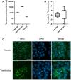
Membrane localization of transferred and transfected ETB-mNG. 0.5 µM ETB-mNG/NDs were incubated with HEK293T cells for 24 h. ETB-mNG was further synthesized in transfected HEK293T cells. Fluorescence microscopy images of fixed cells were taken after 24 h and fluorescence in the cellular membrane was quantified. (A) Comparison of membrane fluorescence in transfected and transferred cells. Mean ± SEM is shown. n = 20 cells. (B) Variation of mean-normalized membrane fluorescence. (C) Representative images of HEK293T cells with transferred ETB-mNG or with synthesized ETB-mNG after transfection.
FIGURE 9
FIGURE 9
Analysis of cluster formation of transferred ETB-mNG. ETB-mNG/ND particles were purified either (i) via the C-terminal Strep-tag of the receptor, (ii) via the Strep-tag and subsequent SEC, or (iii) via LAC by using immobilized 4Ala-ET-1-biotin. ETB-mNG was then transferred into HEK293T cells at standard conditions and representative fluorescence images of the cells were taken. Lower panel: Effects of addition of the endocytosis inhibitors ES9-17 (30 µM) or sucrose (0.45 M) and of performing the transfer at 4°C on the cluster formation of Strep-purified ETB-mNG.
FIGURE 10
FIGURE 10
Co-localization of different MP clusters in transferred HEK293T cells. HEK293T cells were co-incubated with PR-mNG/NDs and ETB-mR3/NDs at standard conditions. After washing and fixation, cells were analyzed by fluorescence microscopy. Images of a representative cell are shown.
FIGURE 11
FIGURE 11
Ligand binding of transferred ETB-mNG in HEK293T cells. LAC-purified ETB-mNG was transferred into HEK293T cells at standard conditions. After washing, cells were incubated with 100 nM DY647-ET-1 for 1 h at 4°C. After additional washing, fluorescence microscopy was performed and representative pictures were taken. Transfected HEK293T cells synthesizing ETB-mNG were used as a positive control.
FIGURE 12
FIGURE 12
Induced internalization of transferred ETB-mNG by cET-1. LAC-purified ETB-mNG was transferred into HEK293T cells at standard conditions. Cells were washed and subsequently incubated with 500 nM cET-1 for 1 h at 4°C. Afterward, they were further incubated at 37°C for 1 h, fixed and ETB-mNG fluorescence in the cytoplasm was quantified via ImageJ. (A) Quantification of ETB-mNG in the cytoplasm of transferred HEK293T cells. Mean ± SEM is shown. n = 20 cells (p < 0.05; paired t-test). (B) Representative images of HEK293T cells treated with or without cET-1 after ETB-mNG transfer.
FIGURE 13
FIGURE 13
Interactions of transferred and transfected GPCRs. HEK293T cells were transfected with constructs expressing either GPRC5B-Myc (A) or HA-IP (B). After 24 h, the transfected cells were incubated with 0.5 µM of either GPRC5B/NDs, IP/NDs, ETB/NDs or empty NDs for 16 h to allow efficient MP transfer. The cells were then washed, lysed and pulldowns based on the Strep-tags of the transferred GPCRs were performed. The MPs were subsequently analyzed for co-purification by SDS-PAGE and immunoblotting. (A) Analysis of transfected cells synthesizing GPRC5B-Myc. Upper panel input: Immunoblotting of cell lysates showing successful expression of GPRC5B-Myc (α-Myc) and the successful transfer of either GPRC5B, IP or ETB (α-Strep). Lower panel Strep-pulldown: Immunoblotting of the eluate from the Strep-column detecting transferred GPCRs (α-Strep) and co-purified transfected GPRC5B-Myc (α-Myc). No pulldown of GPRC5B-Myc was observed with transferred ETB or empty NDs. (B) Analysis of transfected cells synthesizing HA-IP. Upper panel input: Immunoblotting of cell lysates with anti-HA antibody shows a successful expression of HA-IP (α-HA) and the successful transfer of either GPRC5B, IP, or ETB into the cells (α-Strep). Lower panel Strep-pulldown: Immunoblotting of the eluate from the Strep-column detecting transferred GPCRs (α-Strep) and co-purified transfected HA-IP (α-HA).

Similar articles

Cited by

References

    1. Carney C. E., Lenov I. L., Baker C. J., MacRenaris K. W., Eckermann A. L., Sligar S. G., et al. (2015). Nanodiscs as a modular platform for multimodal MR-optical imaging. Bioconjug Chem. 26, 899–905. 10.1021/acs.bioconjchem.5b00107 - DOI - PMC - PubMed
    1. Carvalho J., Chennupati R., Li R., Günther S., Kaur H., Zhao W., et al. (2020). Orphan G protein-coupled receptor GPRC5B controls smooth muscle contractility and differentiation by inhibiting prostacyclin receptor signaling. Circulation 141, 1168–1183. 10.1161/circulationaha.119.043703 - DOI - PubMed
    1. Chau C., Actis P., Hewitt E. (2020). Methods for protein delivery into cells: From current approaches to future perspectives. Biochem. Soc. Trans. 48, 357–365. 10.1042/bst20190039 - DOI - PubMed
    1. Chen W., Smeekens J. M., Wu R. (2016). Systematic study of the dynamics and half-lives of newly synthesized proteins in human cells. Chem. Sci. 7, 1393–1400. 10.1039/c5sc03826j - DOI - PMC - PubMed
    1. Chien C.-T. H., Helfinger L. R., Bostock M. J., Solt A., Tan Y. L., Nietlispach D. (2017). An adaptable phospholipid membrane mimetic system for solution NMR studies of membrane proteins. J. Am. Chem. Soc. 139, 14829–14832. 10.1021/jacs.7b06730 - DOI - PMC - PubMed