

Intravenous Leiomyomatosis with Intracardiac Extension as a Rare Cause of Abdominal Pain in an Adult Patient: A Case Report
- PMID: 35935552
- PMCID: PMC9308881
- DOI: 10.18502/jthc.v16i4.8605
Intravenous Leiomyomatosis with Intracardiac Extension as a Rare Cause of Abdominal Pain in an Adult Patient: A Case Report
Abstract
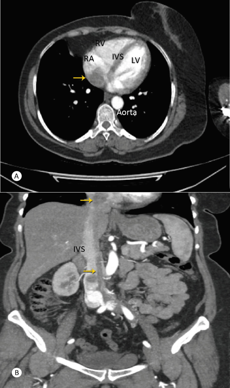
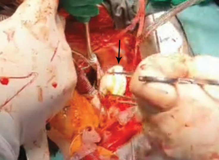
Intravenous leiomyomatosis (IVL) is a rare and benign smooth muscle tumor that arises from intrauterine venules or the myometrium. We herein describe a 49-year-old woman with a history of myomectomy who developed abdominal pain. An intravascular mass with extension to the right atrium was detected in the inferior vena cava. The mass was surgically resected in a single stage under cardiopulmonary bypass. IVL features were indicated by subsequent histopathology. Postoperatively, the patient was diagnosed with massive pericardial effusion and treated with a pericardial window. At 3 months' outpatient clinical follow-up, she was asymptomatic. This case indicates that the diagnosis of IVL with extension to the heart should be kept in mind in patients presenting with abdominal pain.
Keywords: Heart neoplasm, Adult; Leiomyomatosis.
Copyright © 2021 Tehran University of Medical Sciences. Published by Tehran University of Medical Sciences.
Figures

Similar articles
-
Multidisciplinary approach to pelvic leiomyomatosis with intracaval and intracardiac extension: A case report and review of the literature.Gynecol Oncol Rep. 2022 Feb 26;40:100946. doi: 10.1016/j.gore.2022.100946. eCollection 2022 Apr. Gynecol Oncol Rep. 2022. PMID: 35265743 Free PMC article.
-
Massive pelvic recurrence of uterine leiomyomatosis with intracaval-intracardiac extension: video case report and literature review.BMC Surg. 2017 Nov 29;17(1):118. doi: 10.1186/s12893-017-0306-y. BMC Surg. 2017. PMID: 29187188 Free PMC article. Review.
-
"Evolution" of intravascular leiomyomatosis.BMC Womens Health. 2023 Sep 11;23(1):483. doi: 10.1186/s12905-023-02618-3. BMC Womens Health. 2023. PMID: 37697329 Free PMC article.
-
Huge Intravascular Tumor Extending to the Heart: Leiomyomatosis.Case Rep Surg. 2015;2015:658728. doi: 10.1155/2015/658728. Epub 2015 May 31. Case Rep Surg. 2015. PMID: 26114006 Free PMC article.
-
Intracardiac extension of intravenous leiomyomatosis in a pregnant woman: A case report and review of the literature.Can J Cardiol. 2000 Jan;16(1):73-9. Can J Cardiol. 2000. PMID: 10653936 Review.
References
-
- Wang L, He YY, Ren XD, Zhang JY. Intravenous leiomyomatosis involving the right atrium: a case report. Asian J Surg. 2021;44:904–905. - PubMed
-
- Kommoss F, Ebel T, Drusenheimer J, Schelzig H, Lichtenberg A, Fehm T, Aubin H. Die intravenöse leiomyomatose [Intravenous leiomyomatosis] Pathologe. 2019;40:80–84. - PubMed
Publication types
LinkOut - more resources
Full Text Sources