. 2022 Jul 29:2022:1328542.
doi: 10.1155/2022/1328542. eCollection 2022.

The Activation of the Tumor Suppressor Protein p53 by Acriflavine Leads to Mitochondrial Dysfunction and Improves the Radiosensitivity of Colon Cancer Cells

Affiliations

The Activation of the Tumor Suppressor Protein p53 by Acriflavine Leads to Mitochondrial Dysfunction and Improves the Radiosensitivity of Colon Cancer Cells

Caizhao Lin et al. J Immunol Res. .

Abstract

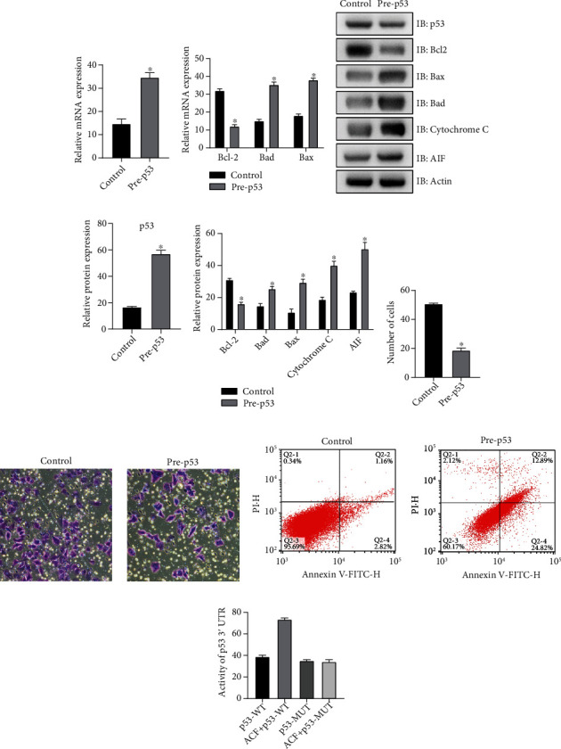
Colon cancer ranks third worldwide, and it has a growing incidence with urbanization and industrialization. Drug resistance in colon cancer is gradually affecting the treatment. This study focused on the mechanisms by which acriflavine (ACF) enhances the radiosensitivity of colon cancer cells. First, the expression and activation levels of tumor suppressor protein p53 were shown high in normal cells and tissues in its detection, which suggests that p53 is likely to be a key factor in colon cancer. Then, the expression of p53 ended up increasing in ACF group after SW620 cells were cultured with ACF. In addition, ACF group had some other changes. The expression of mitochondrial related antiapoptotic protein Bcl-2 increased, while the expression of proapoptotic protein Bax, Bad, cytopigment C, and apoptotic inducer AIF decreased. At the same time, the ability of apoptosis was enhanced, and the ability of proliferation and invasion was decreased. This suggests that ACF can promote p53 expression and affect mitochondrial function and the radiosensitivity of SW620. The luciferase reporting experiment showed that there was a binding site between ACF and p53. Besides, when IR treatment was applied to SW620 with high p53 expression, there was an increase in the expression of Bcl-2 in SW620 and decrease in Bax, Bad, and cytopigment C in AIF. Meanwhile, the cell apoptosis became stronger, and the proliferation and invasion became weaker. The experimental results were similar to those of SW620 cells cultured with ACF, suggesting that p53 is an intermediate factor in the regulation of SW620 by ACF. Finally, in this study, cells were cultured with ACF, and p53 was knocked down at the same time. The experimental results showed that after p53 was knocked down. ACF's ability to regulate SW620 is partially removed. This confirms the view that ACF regulates SW620 cells by regulating p53. In summary, this study found the mechanism by which ACF causes mitochondrial dysfunction and improves the radiosensitivity of colon cancer cells by activating the tumor suppressor protein p53, which may contribute to solving the drug resistance in colon cancer.


Conflict of interest statement

The authors declare that they have no conflicts of interest.

Figures

Figure 1
Figure 1
Low expression of p53 in colon cancer tissues and cells. (a) p53 expression was low in colon cancer cells by QRT-PCR. (b) Low expression of p53 in colon cancer cells by western blot. (c) Low expression of p53 in colon cancer tissues. ∗P < 0.15, compare with the CCD-18co group.
Figure 2
Figure 2
ACF promotes the expression and activation of p53, affects the mitochondrial function of colon cancer cells, and enhances radiosensitivity. (a) The expression of p53 mRNA was increased after ACF treatment. (b) The expression level of p53 became higher after ACF treatment. (c) After ACF treatment, there was a lower the mRNA expression of Bcl-2 and higher mRNA expression levels of Bad and Bax. (d) After ACF treatment, the expression of Bcl-2 decreased, and the expressions of Bad, Bax, AIF, and cytochrome C increased. (e) The proliferation ability of SW620 cells was weakened. (f) The invasion ability of SW620 cells was inhibited. (g) Apoptosis of SW620 cells was enhanced. ∗P < 0.15, compare with the control group. #P < 0.15, compare with the IR group.
Figure 3
Figure 3
Overexpression of p53, mitochondrial dysfunction, and enhanced radiosensitivity of SW620 cells. (a) Overexpression of p53 was detected successfully by QRT-PCR. (b) The experimental results of QRT-PCR showed that the mRNA expression levels of Bcl-2 decreased with overexpression of p53, while the mRNA expression levels of Bad and Bax increased. (c, d) Western Blot results showed that overexpression of p53 led to a decrease in the expression level of Bcl-2, while the expressions of Bad, Bax, AIF, and cytochrome C increased. (e) MTT results showed that overexpression of p53 weakened the proliferation ability of SW620 cells. (f) The results of in vitro invasion experiment showed that the invasion ability of SW620 cells was inhibited by overexpression of p53. (g) Flow cytometry showed that apoptosis of SW620 cells was enhanced. (h) Luciferase reporting experiment results showed that there was a binding site between p53 and ACF. ∗P < 0.15, compare with the control group.
Figure 4
Figure 4
Inhibition of p53 expression, the regulatory effect of ACF on SW620 cells was partially relieved. (a) Western blot results showed that after p53 deletion, the expression level of Bcl-2 increased, while the expressions of Bad, Bax, AIF, and cytochrome C decreased. (b) The experimental results of QRT-PCR showed that the regulation of Bcl-2, Bad, Bax, AIF, and cytochrome C was partially relieved by ACF after p53 was knocked down. (c) MTT test results showed that cell proliferation increased after p53 deletion. (d) The results of in vitro invasion experiment showed that the invasion ability of cells recovered after p53 deletion. (e) Flow cytometry results showed that the apoptosis ability of cells was weakened after p53 deletion. ∗P < 0.15, compare with the control group. #P < 0.15, compare with the ACF + si − p53 group.

Similar articles

Cited by

References

    1. Parkin D. M., Bray F., Ferlay J., Pisani P. Global cancer statistics, 2002. CA: a Cancer Journal for Clinicians . 2005;55(2):74–108. doi: 10.3322/canjclin.55.2.74. - DOI - PubMed
    1. Labianca R., Beretta G. D., Kildani B., et al. Colon cancer. Critical Reviews in Oncology/Hematology . 2010;74(2):106–133. doi: 10.1016/j.critrevonc.2010.01.010. - DOI - PubMed
    1. Berrino F., De Angelis R., Sant M., et al. Survival for eight major cancers and all cancers combined for European adults diagnosed in 1995-99: results of the EUROCARE-4 study. The Lancet Oncology . 2007;8(9):773–783. doi: 10.1016/S1470-2045(07)70245-0. - DOI - PubMed
    1. Kwak E. L., Chung D. C. Hereditary colorectal cancer syndromes: an overview. Clinical Colorectal Cancer . 2007;6(5):340–344. doi: 10.3816/CCC.2007.n.002. - DOI - PubMed
    1. Park S. Y., Kim J. T., Park E. S., et al. Collapsin response mediator protein 4 enhances the radiosensitivity of colon cancer cells through calcium-mediated cell signaling. Oncology Reports . 2021;45(4):p. 6. doi: 10.3892/or.2021.7957. - DOI - PMC - PubMed

MeSH terms

Substances