Review
. 2022 Jul 22:9:961703.
doi: 10.3389/fmed.2022.961703. eCollection 2022.

Role of gut microbiota-derived signals in the regulation of gastrointestinal motility

Affiliations
Review

Role of gut microbiota-derived signals in the regulation of gastrointestinal motility

Zhipeng Zheng et al. Front Med (Lausanne). .

Abstract

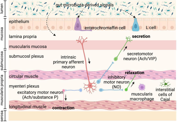
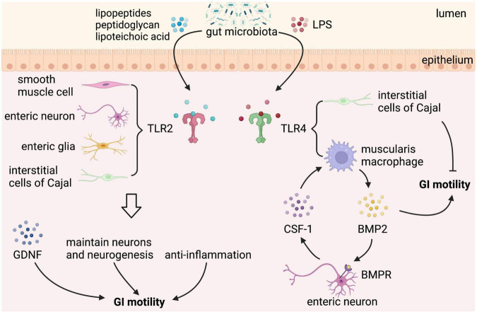
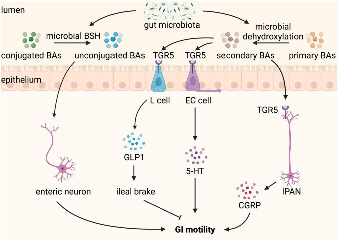
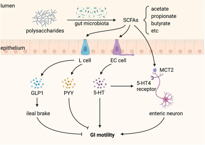
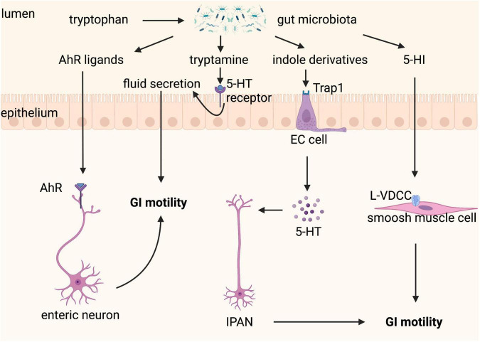
The gastrointestinal (GI) tract harbors trillions of commensal microbes, called the gut microbiota, which plays a significant role in the regulation of GI physiology, particularly GI motility. The GI tract expresses an array of receptors, such as toll-like receptors (TLRs), G-protein coupled receptors, aryl hydrocarbon receptor (AhR), and ligand-gated ion channels, that sense different gut microbiota-derived bioactive substances. Specifically, microbial cell wall components and metabolites, including lipopeptides, peptidoglycan, lipopolysaccharides (LPS), bile acids (BAs), short-chain fatty acids (SCFAs), and tryptophan metabolites, mediate the effect of gut microbiota on GI motility through their close interactions with the enteroendocrine system, enteric nervous system, intestinal smooth muscle, and immune system. In turn, GI motility affects the colonization within the gut microbiota. However, the mechanisms by which gut microbiota interacts with GI motility remain to be elucidated. Deciphering the underlying mechanisms is greatly important for the prevention or treatment of GI dysmotility, which is a complication associated with many GI diseases, such as irritable bowel syndrome (IBS) and constipation. In this perspective, we overview the current knowledge on the role of gut microbiota and its metabolites in the regulation of GI motility, highlighting the potential mechanisms, in an attempt to provide valuable clues for the development of gut microbiota-dependent therapy to improve GI motility.

Keywords: bile acid; gastrointestinal motility; gut microbial components; gut microbiota; short-chain fatty acids (SCFAs); tryptophan metabolites.


Conflict of interest statement

The authors declare that the research was conducted in the absence of any commercial or financial relationships that could be construed as a potential conflict of interest.

Figures

FIGURE 1
FIGURE 1
Anatomy of the bowel wall ensures the effect of gut microbiota on gastrointestinal (GI) motility. Gut microbiota is geographically close to the bowel wall, which is composed of the mucosa layer (epithelium, lamina propria, and muscularis mucosa), the submucosa layer (submucosal plexus), the muscularis propria (circular smooth muscle, myenteric plexus, and longitudinal smooth muscle), and the serosa layer. Enteroendocrine cells (enterochromaffin cells and L cells) dispersed among the mucosa layer can directly sense gut microbiota-derived signals and then secrete hormones, such as glucagon-like peptides (GLPs) and peptide YY (PYY) (L cells), and serotonin (enterochromaffin cells), affecting enteric nervous system (ENS) and gastrointestinal (GI) motility. The ENS comprising submucosal plexus and myenteric plexus plays a central role in GI motility and can also sense and respond to gut microbiota-derived stimuli that cross the epithelium. The myenteric plexus is responsible for the propulsion of intestinal contents under the movement of the smooth muscle, while the submucosal plexus is mainly involved in the secretion and absorption. Intrinsic primary afferent neurons (IPANs) are activated by gut-derived signals and activate ascending and descending interneurons, which stimulate inhibitory and excitatory motor neurons, as well as secretomotor neurons. Besides, musculari macrophage and interstitial cells of Cajal (ICCs) in muscularis propria can be activated by gut microbiota-derived signals affecting GI motility. Ach, acetylcholine; NO, nitric oxide; VIP, vasoactive intestinal peptide.
FIGURE 2
FIGURE 2
Gastrointestinal (GI) motility is highly dependent on gut microbiota. Both antibiotic-treated and germ-free (GF) rodents that lack gut microbiota have slowed GI motility, with prolonged intestinal transit, attenuated contractility, reduced defecation frequency, and loss of enteric neurons. Probiotic supplements, such as Lactobacillus acidophilus, Bifidobacterium bifidum, and Akkermansia. muciniphila can improve GI motility.
FIGURE 3
FIGURE 3
Gut microbial components regulate gastrointestinal (GI) motility via binding to toll-like receptor 2/4. Toll-like receptors (TLRs) expressed in the GI tract sense gut microbial components take part in the regulation of GI motility. TLR2 is expressed on enteric smooth muscle cells, neurons, neuroglia, and interstitial cells of Cajal (ICCs). Lipopeptides, peptidoglycan, and lipoteichoic acid from gut microbiota binding to TLR2 stimulate the release of glial cell line-derived neurotrophic factor (GDNF), maintain neurons and neurogenesis, and play an anti-inflammation effect, which can improve GI motility. In addition to TLR2, TLR4 is the best-characterized receptor recognizing gut microbiota-derived Lipopolysaccharide (LPS). LPS binding to TLR4 expressed on muscularis macrophage (MM) stimulates the release of bone morphogenetic protein 2 (BMP2), which improves GI motility. In response to BMP2, enteric neurons produce colony stimulatory factor 1 (CSF-1), which in turn promotes MM homeostasis. However, LPS binding to TLR4 expressed on ICCs has a negative effect on GI motility.
FIGURE 4
FIGURE 4
Bile acids (BAs) converted by gut microbiota regulate gastrointestinal (GI) motility. BAs are produced from cholesterol in the liver and released into the GI tract when food intake. In the GI tract, conjugated BAs can be converted by microbial bile salt hydrolase (BSH) to unconjugated BAs, which stimulate enteric neurons and promote GI motility. Primary BAs can be converted by microbial dehydroxylation to secondary BAs, which binding to Taketa G-protein-coupled receptor 5 (TGR5) expressed L cell and enterochromaffin (EC) cell stimulate the release of glucagon-like peptide 1 (GLP-1) and 5-hydroxytryptamine (5-HT), respectively. GLP1 leads to ileal brake and slows GI motility, whereas 5-HT promotes GI motility. TGR5 is also expressed on intrinsic afferent primary neurons (IPANs), which can be activated by secondary BAs and produce calcitonin gene-related peptide (CGRP) improving GI motility.
FIGURE 5
FIGURE 5
Short-chain fatty acids (SCFAs) produced by gut microbiota regulate gastrointestinal (GI) motility. SCFAs, including acetate, propionate, and butyrate, are produced from gut microbial fermentation of dietary polysaccharides. L cells sense SCFAs and produce glucagon-like peptide 1 (GLP-1) and peptide YY (PYY), both of which inhibit GI motility. Enterochromaffin (EC) cells sense SCFAs and produce 5-hydroxytryptamine (5-HT), which promotes GI motility by activating the 5-HT4 receptor expressed on enteric neurons. SCFAs can also stimulate enteric neurons through monocarboxylate transporter 2 (MCT2), playing a positive role in GI motility.
FIGURE 6
FIGURE 6
Tryptophan metabolism controlled by gut microbiota regulates gastrointestinal (GI) motility. Tryptophan can be metabolized by gut microbiota to a variety of active substances. Aryl hydrocarbon receptor (AhR) ligands binding to AhR expressed on enteric neurons promote GI motility. Tryptamine contributes to fluid secretion by activating the 5-hydroxytryptamine (5-HT) receptor on enterocytes, which increases GI motility. Indole derivatives stimulate the release of 5-HT from enterochromaffin (EC) cells via transient receptor potential ankyrin A1 (Trpa1), which improves GI motility through stimulating intrinsic afferent primary neurons (IPANs). 5-hydroxyindole (5-HI) can directly act on smooth muscle cells via L-type voltage-dependent calcium channels (L-VDCCs) and then promotes GI motility.

References

    1. Boeckxstaens G, Camilleri M, Sifrim D, Houghton LA, Elsenbruch S, Lindberg G, et al. Fundamentals of neurogastroenterology: physiology/motility - sensation. Gastroenterology. (2016) S0016-5085:221–3. 10.1053/j.gastro.2016.02.030 - DOI - PubMed
    1. Black CJ, Ford AC. Global burden of irritable bowel syndrome: trends, predictions and risk factors. Nat Rev Gastroenterol Hepatol. (2020) 17:473–86. 10.1038/s41575-020-0286-8 - DOI - PubMed
    1. Ford AC, Sperber AD, Corsetti M, Camilleri M. Irritable bowel syndrome. Lancet. (2020) 396:1675–88. 10.1016/S0140-6736(20)31548-8 - DOI - PubMed
    1. Vriesman MH, Koppen IJN, Camilleri M, Lorenzo C, Di, Benninga MA. Management of functional constipation in children and adults. Nat Rev Gastroenterol Hepatol. (2020) 17:21–39. 10.1038/s41575-019-0222-y - DOI - PubMed
    1. Wattchow D, Heitmann P, Smolilo D, Spencer NJ, Parker D, Hibberd T, et al. Postoperative ileus-an ongoing conundrum. Neurogastroenterol Motil. (2021) 33:e14046. 10.1111/nmo.14046 - DOI - PubMed

LinkOut - more resources