

Retinal and choroidal microvascular alterations in Behcet's disease without ocular manifestations: A systematic review and meta-analysis
- PMID: 35935767
- PMCID: PMC9353174
- DOI: 10.3389/fmed.2022.911990
Retinal and choroidal microvascular alterations in Behcet's disease without ocular manifestations: A systematic review and meta-analysis
Abstract
Purpose: We performed a systematic review and meta-analysis to examine the microvascular alterations in non-ocular Behcet's disease (BD) using optical coherence tomography angiography (OCTA).
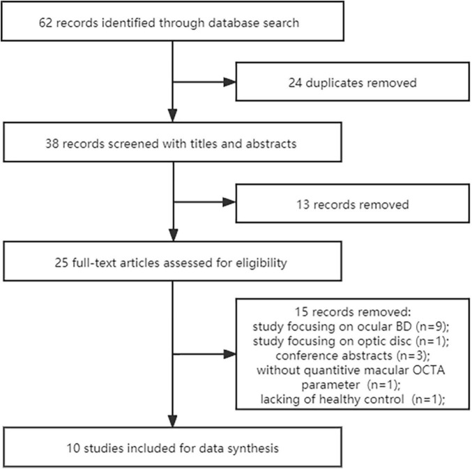
Methods: A comprehensive search was performed in Pubmed, Embase and Cochrane databases for eligible studies from inception to February 2022. Detailed clinical demographics were extracted from each study by two independent reviewers. The weighted mean difference (WMD) and 95% confidence intervals (CI) were used to compare the OCTA parameters between non-ocular BD and healthy controls. Stata 12.0 was adopted to conduct statistical analyses.
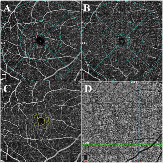
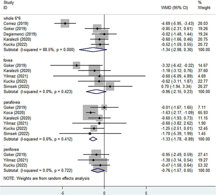
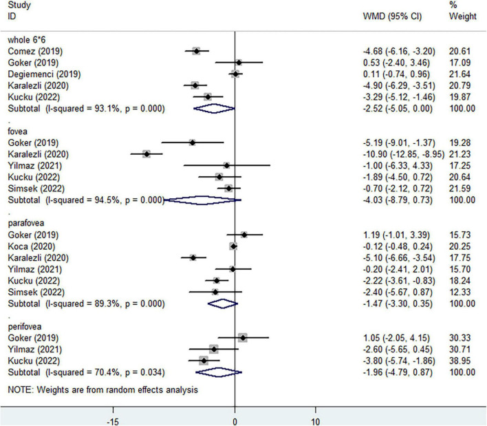
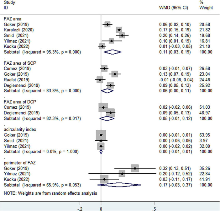
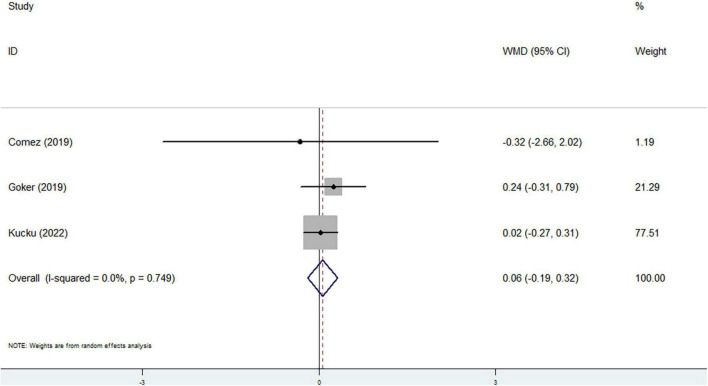
Results: Ten cross-sectional studies involving 386 eyes in non-ocular BD and 418 eyes in healthy volunteers were ultimately included in the present analysis. When considering superficial capillary plexus (SCP) and deep capillary plexus (DCP), no significant differences of vessel densities in the whole enface image, fovea and perifovea were evaluated between two groups. Significantly reduced parafoveal vessel density of SCP was observed in non-ocular BD in comparison with healthy group (WMD = -1.33, 95%CI: -1.78, -0.89; I 2 = 0.6%), while slightly decreased parafoveal vessel density was assessed in DCP (WMD = -1.47, 95%CI: -3.30, 0.35; I 2 = 89.3%). Significantly increasing foveal avascular zone (FAZ) area was observed in non-ocular BD when compared to healthy controls (WMD = 0.11, 95%CI: 0.03, 0.19; I 2 = 95.3%). There was no significant difference in flow area of choriocapillaris between non-ocular BD and control group (WMD = 0.06, 95%CI: -0.19, 0.32; I 2 = 0%).
Conclusion: Based on current analysis, our results demonstrated significantly lower parafoveal vessel density of SCP and lager FAZ area in full vasculature in non-ocular BD. The retinal microvascular alterations appear before the emergence of ocular manifestations.
Systematic trial registration: [https://www.crd.york.ac.uk/PROSPERO/], identifier [CRD42021244856].
Keywords: Behcet’s disease; macula; meta-analysis; optical coherence tomography angiography (OCTA); vessel density.
Copyright © 2022 Fan, Shi, Chen, Li, Yu and Li.
Conflict of interest statement
The authors declare that the research was conducted in the absence of any commercial or financial relationships that could be construed as a potential conflict of interest.
Figures
References
-
- Lancet. Criteria for diagnosis of Behçet’s disease. International study group for Behçet’s disease. Lancet. (1990) 335:1078–80. - PubMed
Publication types
LinkOut - more resources
Full Text Sources
Miscellaneous