. 2022 Jul 22:13:919940.
doi: 10.3389/fphar.2022.919940. eCollection 2022.

Nitidine Chloride Alleviates Inflammation and Cellular Senescence in Murine Osteoarthritis Through Scavenging ROS

Affiliations

Nitidine Chloride Alleviates Inflammation and Cellular Senescence in Murine Osteoarthritis Through Scavenging ROS

Changjian Lin et al. Front Pharmacol. .

Abstract

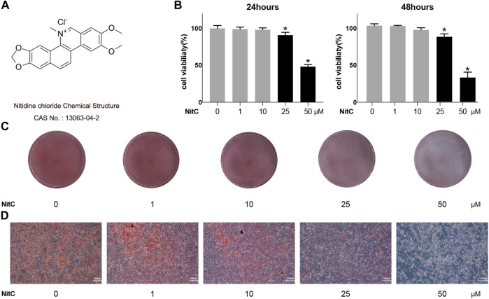
Osteoarthritis (OA) is one of the most common chronic musculoskeletal disorder worldwide, representing a major source of disability, pain and socioeconomic burden. Yet the effective pharmaceutical treatments applied in the clinical works are merely symptomatic management with uncertainty around their long-term safety and efficacy, namely no drugs currently are capable of modulating the biological progression of OA. Here, we identified the potent anti-inflammatory as well as anti-oxidative properties of Nitidine Chloride (NitC), a bioactive phytochemical alkaloid extracted from natural herbs, in IL-1β-treated rat articular chondrocytes (RACs), LPS-stimulated RAW 264.7 and rat osteoarthritic models in vivo. We demonstrated NitC remarkably inhibited the production of inflammatory mediators including COX2 and iNOS, suppressed the activation of MAPK and NF-κB cell signaling pathway and reduced the expression of extracellular matrix (ECM) degrading enzymes including MMP3, MMP9 and MMP13 in IL-1β-treated RACs. Several emerging bioinformatics tools were performed to predict the underlying mechanism, the result of which indicated the potential reactive oxygen species (ROS) clearance potential of NitC. Further, NitC exhibited its anti-oxidative potential through ameliorating cellular senescence in IL-1β-treated RACs and decreasing NLRP3 inflammasomes activation in LPS-stimulated RAW 264.7 via scavenging ROS. Additionally, X-ray, micro-CT and other experiments in vivo demonstrated that intra-articular injection of NitC significantly alleviated the cartilage erosion, ECM degradation and subchondral alterations in OA progression. In conclusion, the present study reported the potent anti-inflammatory and anti-oxidative potential of NitC in OA biological process, providing a promising therapeutic agent for OA management.

Keywords: MAPK; NF-κB; NLRP3 inflammasome; ROS; nitidine chloride; osteoarthritis; senescence.


Conflict of interest statement

The authors declare that the research was conducted in the absence of any commercial or financial relationships that could be construed as a potential conflict of interest.

Figures

FIGURE 1
FIGURE 1
Effect of NitC on viability and chondrocyte phenotype maintenance. (A) Nitidine Chloride (NitC) chemical structure. (B) CCK8 analysis of NitC on chondrocytes viability. (C) Gross view of safranin O stained primary rat articular chondrocytes treated with 0, 1, 10, 25, or 50 μM NitC in 12 wells for 24 h. (D) Microscopic images of safranin O stained primary rat chondrocytes treated with 0, 1, 10, 25, or 50 μM NitC for 24 h. *p < 0.05 versus the control group. Scale bar = 200 μM.
FIGURE 2
FIGURE 2
NitC alleviated ECM degradation and inhibited RACs inflammation in vitro. (A) The WB analysis of COL2, iNOS, MMP9, COX2, MMP13, MMP3, and GA was conducted and their relative protein expression (B) was shown. (C–G) The qRT-PCR analysis was performed to detect the relative mRNA expression of iNOS, MMP9, MMP13, COX2, and COL2. (H) Representative images of the Safranin O staining of pretreated chondrocytes. (I) The microscopic images of Alcian Blue staining of pretreated chondrocytes. #p < 0.05 versus the control group. ∗p < 0.05 versus the model group. Scale bars = 200 μM.
FIGURE 3
FIGURE 3
NitC inhibited MAPK and NF-κB pathway in the osteoarthritic model in vitro. (A) The WB analysis of MAPK pathway-related protein: p-ERK, ERK, p-JNK, JNK, p-p38, and p38 as well as endogenous control GA were performed as previously described. Besides, the p-ERK/ERK, p-JNK/JNK, and p-p38/p38 ratios were shown. (B) The WB analysis of NF-κB pathway-related protein: p-p65, p65, p-IκBα, IκBα, and endogenous control β-Actin were conducted. The p-p65/p65 and p-IκBα/IκBα ratios were shown. (C) The nuclear translocation of p65 was assessed by immunofluorescent microscopy. The quantified results of nuclear location/total ratio were shown as well. Blue: DAPI. Green: p65. #p < 0.05 versus the control group. ∗p < 0.05 versus the model group. Scale bars = 100 μM.
FIGURE 4
FIGURE 4
NitC reduced ROS generation and ameliorated cellular senescence in RACs. (A) The potential targets of NitC were obtained by PharmMapper. The protein-protein association network of the targets was established by STRING. (B) The top 10 hub genes were calculated by Cytoscape subsequently. Genes marked with “ROS” are involved with the modulation of ROS generation according to the existing research. (C) Immunofluorescent microscopy was utilized to detect the ROS in RACs. The intensity of fluorescence represented the relative level of ROS. (D) The ROS levels of different groups were also assessed through flow cytometry. (E) β-Galactosidase activity assay was performed in RACs. Typical normal RAC and senescent RAC were shown. Red arrows mark the senescent RAC. (F) The WB analysis of cellular senescence-related genes: p16 and p53 were conducted. Meanwhile, the quantified results of relative protein expression were present. #p < 0.05 versus the control group. ∗p < 0.05 versus the model group. Scale bars = 50 μM.
FIGURE 5
FIGURE 5
Regeneration of ROS rescue the impact of NitC on RACs. (A) The ROS levels in RACs were detected through immunofluorescence analysis. (B) β-Galactosidase activity assay was performed to identify the senescent RACs. (C) WB analysis of iNOS, COX2, MMP9, and β-Actin. The relative protein expression was quantified and shown respectively in (D–F). ∗p < 0.05 versus the model group, other significant differences between the groups were shown as indicated in the figure.
FIGURE 6
FIGURE 6
NitC inhibited NLRP3-inflammasome activation through scavenging ROS. (A) The WB analysis and quantitative analysis of the (B) cleaved IL-1β, (C) pro-IL-1β, (D) NLRP3, (E) COX2, and (F) iNOS. (G) Another WB analysis was performed to investigate the role of ROS in NitC-treated RAW 264.7. The quantified results of (H) pro-IL-1β, (I) COX2, (J) NLRP3, and (K) iNOS were shown in the figure. ∗p < 0.05 versus the model group, other significant differences between the groups were shown as indicated in the figure.
FIGURE 7
FIGURE 7
Intra-articular injection of NitC ameliorated OA progression in rat OA model. (A) The flowchart for animal procedures. (B) The resected medial meniscus. (C) Microscopic images of safranin O staining of rat knee joints. (D) Representative images of HE staining of rat knee joints. (E) Typical images of immunofluorescence analysis against type II collagen and MMP13. Scale bars = 200 μM.
FIGURE 8
FIGURE 8
Radiological assessment of NitC impact on DMM rats. (A) Representative images of rat knees X-ray scanning. The treatment was performed as previously described in Methods and Materials. (B) The coronal plane of rat knees. The red arrow showed osteophyte formation. (C) Subchondral bone in each group. (D) Reconstruction of 3D rat knee models. IMALYTICS Preclinical 2.1 software was used to evaluate BV/TV (E), Tb.Th (F), and Tb.Sp (G). #p < 0.05 versus the control group. ∗p < 0.05 versus the model group.

Similar articles

Cited by

References

    1. Ansari M. Y., Ahmad N., Haqqi T. M. (2020). Oxidative Stress and Inflammation in Osteoarthritis Pathogenesis: Role of Polyphenols. Biomed. Pharmacother. 129, 110452. 10.1016/j.biopha.2020.110452 - DOI - PMC - PubMed
    1. Au M., Liu Z., Rong L., Zheng Y., Wen C. (2020). Endothelin-1 Induces Chondrocyte Senescence and Cartilage Damage via Endothelin Receptor Type B in a Post-Traumatic Osteoarthritis Mouse Model. Osteoarthr. Cartil. 28, 1559–1571. 10.1016/j.joca.2020.08.006 - DOI - PubMed
    1. Bai Y., Gong X., Dou C., Cao Z., Dong S. (2019). Redox Control of Chondrocyte Differentiation and Chondrogenesis. Free Radic. Biol. Med. 132, 83–89. 10.1016/j.freeradbiomed.2018.10.443 - DOI - PubMed
    1. Bao J., Lin C., Zhou X., Ma D., Ge L., Xu K., et al. (2021). CircFAM160A2 Promotes Mitochondrial Stabilization and Apoptosis Reduction in Osteoarthritis Chondrocytes by Targeting miR-505-3p and SIRT3. Oxid. Med. Cell. Longev. 2021, 5712280. 10.1155/2021/5712280 - DOI - PMC - PubMed
    1. Bermúdez-Muñoz J. M., Celaya A. M., Hijazo-Pechero S., Wang J., Serrano M., Varela-Nieto I. (2020). G6PD Overexpression Protects from Oxidative Stress and Age-Related Hearing Loss. Aging Cell 19, e13275–18. 10.1111/acel.13275 - DOI - PMC - PubMed