. 2022 Jul 22:13:946192.
doi: 10.3389/fphar.2022.946192. eCollection 2022.

Class IIa histone deacetylase inhibition ameliorates acute kidney injury by suppressing renal tubular cell apoptosis and enhancing autophagy and proliferation

Affiliations

Class IIa histone deacetylase inhibition ameliorates acute kidney injury by suppressing renal tubular cell apoptosis and enhancing autophagy and proliferation

Jialu Li et al. Front Pharmacol. .

Abstract

Expression and function of histone deacetylases (HDACs) vary with cell types and pathological conditions. Our recent studies showed that pharmacological targeting class IIa HDACs attenuated renal fibrosis, but the effect of class IIa HDAC inhibition on acute kidney injury (AKI) remains unknown. In this study, we found that four class IIa HDACs (4, 5, 7, 9) were highly expressed in the kidney of folic acid (FA) and ischemia/reperfusion (I/R)-induced AKI in mice. Administration of TMP269, a potent and selective class IIa HDAC inhibitor, improved renal function and reduced tubular cell injury and apoptosis, with concomitant suppression of HDAC4 and elevation of acetyl-histone H3. Mechanistical studies showed that TMP269 treatment inhibited FA and I/R-induced caspase-3 cleavage, Bax expression and p53 phosphorylation. Conversely, TMP269 administration preserved expression of E-cadherin, BMP7, Klotho and Bcl-2 in injured kidneys. Moreover, TMP269 was effective in promoting cellular autophagy as indicated by increased expression of Atg7, beclin-1, and LC3II, and promoted renal tubular cell proliferation as shown by increased number of proliferating cell nuclear antigen-positive cells and expression of cyclin E. Finally, blocking class IIa HDACs inhibited FA-and I/R-induced phosphorylation of extracellular signal-regulated kinases 1 and 2, and p38, two signaling pathways associated with the pathogenesis of AKI. Collectively, these results suggest that pharmacological inhibition of class IIa HDACs protects against AKI through ameliorating apoptosis, enhancing autophagy and promoting proliferation of renal tubular cells by targeting multiple signaling pathways.

Keywords: TMP269; acute kidney injury; apoptosis; autophagy; class IIa histone deacetylases; folic acid; ischemia/reperfusion; renal tubular cells.


Conflict of interest statement

The authors declare that the research was conducted in the absence of any commercial or financial relationships that could be construed as a potential conflict of interest.

Figures

FIGURE 1
FIGURE 1
Expression of HDAC4, HDAC5, HDAC7, and HDAC9 in the kidney following ischemia/reperfusion (I/R) and folic acid (FA) injury (A) At 48 h after sham operation or I/R, kidneys were collected and tissue lysates were subjected to immunoblot analysis with antibodies against HDAC4, 5, 7, 9, and GAPDH. Representative immunoblots are three samples in each group (B) Expression levels of HDAC4, 5, 7, 9 were quantified by densitometry and normalized with GAPDH. Data are expressed as means ± SD (n = 3). **p < 0.01 (C) At 48 h after sham operation, FA, or I/R, kidneys were collected and tissue sections were subjected to immunohistochemical staining followed by photomicrograph illustrating protein expression of individual class IIa HDACs. Scale bar, 20 µm. Original magnification, ×200.
FIGURE 2
FIGURE 2
Administration of TMP269 improves renal function and attenuates renal pathological changes in the kidney following FA and I/R injury. At 48 h after various treatments as indicated, blood was collected and the serum creatinine and blood urea nitrogen (BUN) were measured (A,B,E,F). The kidneys injured by FA (D) and I/R (H) underwent hematoxylin and eosin (HE) staining. Scale bar, 20 µm. Original magnification, ×200. Morphological changes induced by FA (C) and I/R (G) and were scored based on the scale described in Material and methods (I) After IR, kidney tissue lysates were subjected to immunoblot analysis with specific antibodies against HDAC4, acetyl-histone H3 (acetyl-H3), histone H3 and GAPDH. Expression levels of HDAC4 (J) and acetyl-histone histone 3 (K) were quantified by densitometry and normalized with GAPDH and histone H3, respectively. Data are represented as means ± SD (n = 3). **p < 0.01; ***p < 0.001.
FIGURE 3
FIGURE 3
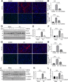
Administration of TMP269 alleviates renal tubular injury and renal tubular cell apoptosis in the kidney following FA and I/R injury. Photomicrographs (×200) illustrate NGAL or TUNAL immunofluorescent staining of the kidney tissues collected at 48 h after FA (A) or I/R (G) injury with or without TMP269 treatment. Scale bar, 20 µm (B,H) Tubules with positive NGAL staining or (C,I) tubular cells with TUNAL staining were counted in 10 high-power fields and expressed as means ± SD, respectively. Scale bar, 20 µm. Original magnification, ×200 (D,J) Kidneys injured by IR or FA, respectively as indicated, kidney tissue lysates were subjected to immunoblot analysis with specific antibodies against NGAL, GAPDH, cleaved caspase-3 (C-caspase-3) or α-tubulin. Expression levels of NGAL (E,K) and cleaved caspase-3 (F,L) were quantified by densitometry and normalized with GAPDH and α-tubulin, respectively. Data are represented as means ± SD (n = 3). **p < 0.01; ***p < 0.001.
FIGURE 4
FIGURE 4
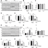
Administration of TMP269 regulates the expression and activation of apoptosis-related signal molecules in the kidney following FA and I/R injury (A,G) Kidneys were injured by FA or IR, respectively, and tissue lysates were subjected to immunoblot analysis with specific antibodies against phospho-p53 (p-p53), p53, Bax, Bcl-2 or β-actin. Representative immunoblots are three samples in each group (B,H) Expression levels of phospho-p53 (p-p53) were quantified by densitometry and normalized with p53 (C–E, I–K) Expression levels of p53, Bax or Bcl-2 were quantified by densitometry, respectively and normalized with b-actin, respectively (F,L) The ratio of Bcl-2/Bax were calculated. Data are means ± SD (n = 3). *<0.05; **p < 0.01; ***p < 0.001.
FIGURE 5
FIGURE 5
Administration of TMP269 enhances autophagy in the kidney following FA and I/R injury (A,E) Kidneys were injured by FA or IR, respectively, and tissue lysates were subjected to immunoblot analysis with specific antibodies against Atg7, Beclin-1, LC3, and GAPDH. Representative immunoblots are three samples in each group. Expression levels of Atg7 (B,F), Beclin-1 (C,G), or LC3 (D,H) were quantified by densitometry and normalized with GAPDH. Data are means ± SD (n = 3). *<0.05; **p < 0.01; ***p < 0.001.
FIGURE 6
FIGURE 6
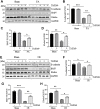
Administration of TMP269 preserves expression of E-cadherin, BMP7 and Klotho in the kidney following FA and I/R injury (A,E) Kidneys were injured by FA or IR, respectively, and tissue lysates were subjected to immunoblot analysis with specific antibodies against E-cadherin, BMP7, Klotho and GAPDH. Representative immunoblots are three samples in each group. Expression levels of E-cadherin (B,F), BMP-7 (C,G), or Klotho (D,H) were quantified by densitometry and normalized with GAPDH. Data are means ± SD (n = 3). *<0.05; **p < 0.01; ***p < 0.001.
FIGURE 7
FIGURE 7
Administration of TMP269 promotes cell proliferation in the kidney following FA and I/R injury (A,F) Photomicrographs (×200) illustrate proliferating cell nuclear antigen (PCNA) immunofluorescent staining of the kidney tissues collected from murine models of FA or I/R-induced AKI. Scale bar, 20 µm. Original magnification, ×200 (B,G) Tubular cells with positive PCNA staining were counted in 10 high-power fields and expressed as means ± SD (C,I) Kidney tissue lysates were subjected to immunoblot analysis with specific antibodies against PCNA, Cyclin E or GAPDH. Representative immunoblots are three samples in each group. Expression levels of PCNA (D,H) and Cyclin E (E,J) were quantified by densitometry and normalized with GAPDH, respectively. Data are means ± SD (n = 3). *<0.05; **p < 0.01; ***p < 0.001.
FIGURE 8
FIGURE 8
Administration of TMP269 inhibits MAPK signaling pathways (A,D) Kidneys were injured by FA or IR, respectively, and tissue lysates were subjected to immunoblot analysis with specific antibodies against p-ERK1/2, ERK1/2, p-p38, p38 or α-tubulin. Representative immunoblots are three samples in each group. Expression levels of p-ERK1/2 (B,E) and p-38 (C,F) were quantified by densitometry and normalized with ERK1/2 and p38, respectively. Data are means ± SD (n = 3). *<0.05; **p < 0.01; ***p < 0.001.

Similar articles

Cited by

References

    1. Agarwal A., Dong Z., Harris R., Murray P., Parikh S. M., Rosner M. H., et al. X. W. G. A cute Dialysis Quality Initiative (2016). Cellular and molecular mechanisms of AKI. J. Am. Soc. Nephrol. 27 (5), 1288–1299. 10.1681/asn.2015070740 - DOI - PMC - PubMed
    1. Archdeacon P., Detwiler R. K. (2008). Bone morphogenetic protein 7 (BMP7): A critical role in kidney development and a putative modulator of kidney injury. Adv. Chronic Kidney Dis. 15 (3), 314–320. 10.1053/j.ackd.2008.04.011 - DOI - PubMed
    1. Brenner B., Weinmann S., Grassmé H., Lang F., Gulbins O., Gulbins E. (1997). L-selectin activates JNK via src-like tyrosine kinases and the small G-protein Rac. Immunology 92 (2), 214–219. 10.1046/j.1365-2567.1997.00336.x - DOI - PMC - PubMed
    1. Cappellacci L., Perinelli D. R., Maggi F., Petrelli M., Petrelli R. (2020). Recent progress in histone deacetylase inhibitors as anticancer agents. Curr. Med. Chem. 27 (15), 2449–2493. 10.2174/0929867325666181016163110 - DOI - PubMed
    1. Costalonga E. C., Silva F. M., Noronha I. L. (2016). Valproic acid prevents renal dysfunction and inflammation in the ischemia-reperfusion injury model. Biomed. Res. Int. 2016, 5985903. 10.1155/2016/5985903 - DOI - PMC - PubMed