. 2022 Jul 21:13:939542.
doi: 10.3389/fphar.2022.939542. eCollection 2022.

Development and validation of a hypoxia-stemness-based prognostic signature in pancreatic adenocarcinoma

Affiliations

Development and validation of a hypoxia-stemness-based prognostic signature in pancreatic adenocarcinoma

Xiong Tian et al. Front Pharmacol. .

Abstract

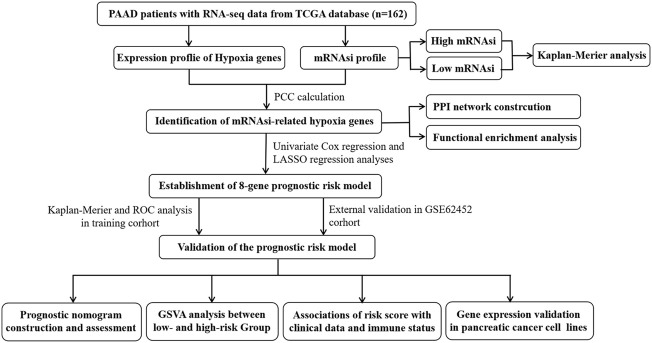
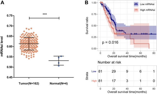
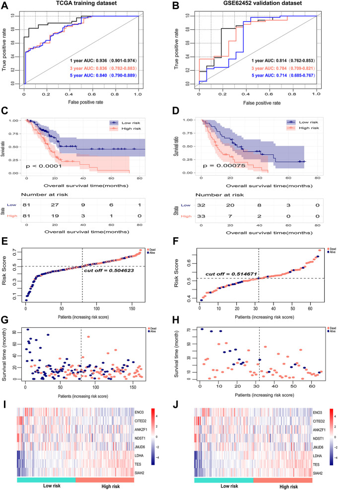
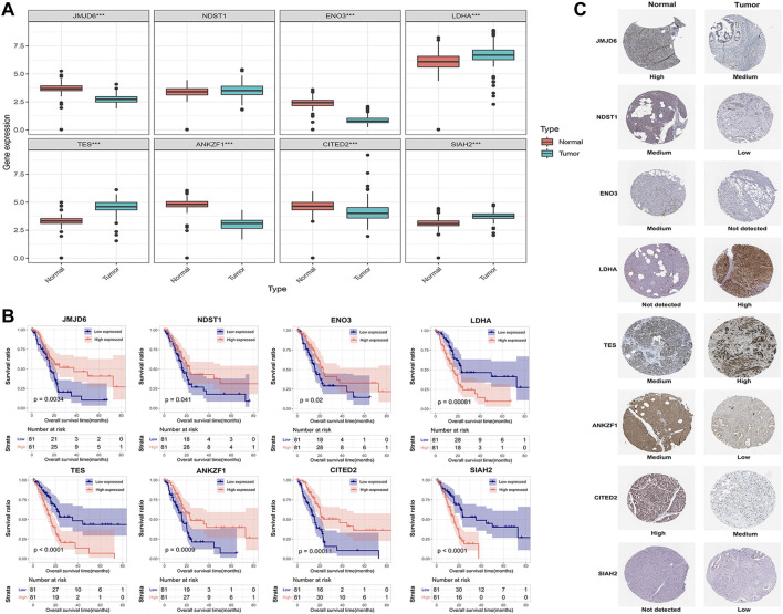
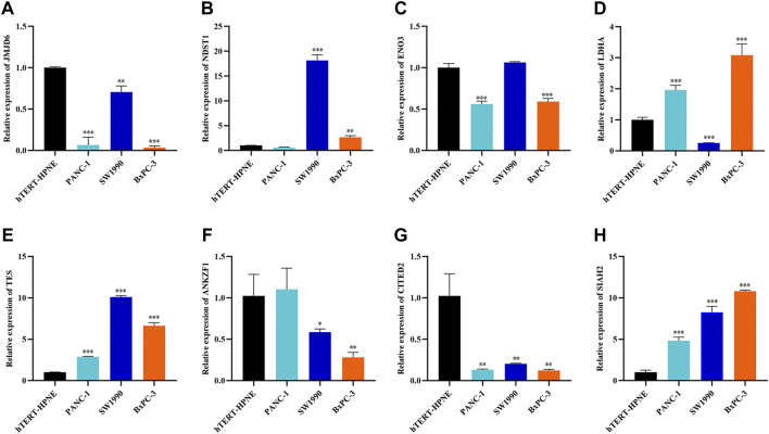
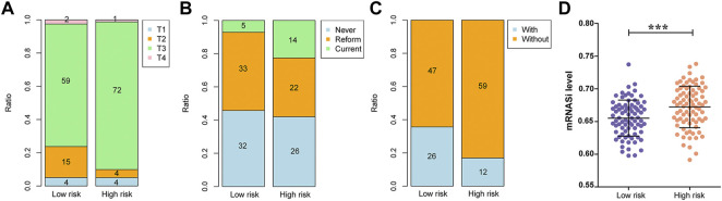
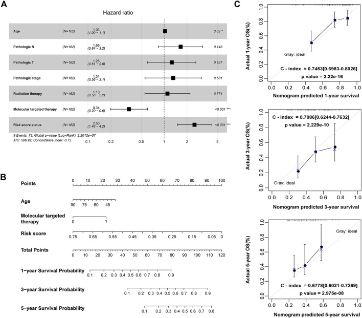
Background: Pancreatic adenocarcinoma (PAAD) is one of the most aggressive and fatal gastrointestinal malignancies with high morbidity and mortality worldwide. Accumulating evidence has revealed the clinical significance of the interaction between the hypoxic microenvironment and cancer stemness in pancreatic cancer progression and therapies. This study aims to identify a hypoxia-stemness index-related gene signature for risk stratification and prognosis prediction in PAAD. Methods: The mRNA expression-based stemness index (mRNAsi) data of PAAD samples from The Cancer Genome Atlas (TCGA) database were calculated based on the one-class logistic regression (OCLR) machine learning algorithm. Univariate Cox regression and LASSO regression analyses were then performed to establish a hypoxia-mRNAsi-related gene signature, and its prognostic performance was verified in both the TCGA-PAAD and GSE62452 corhorts by Kaplan-Meier and receiver operating characteristic (ROC) analyses. Additionally, we further validated the expression levels of signature genes using the TCGA, GTEx and HPA databases as well as qPCR experiments. Moreover, we constructed a prognostic nomogram incorporating the eight-gene signature and traditional clinical factors and analyzed the correlations of the risk score with immune infiltrates and immune checkpoint genes. Results: The mRNAsi values of PAAD samples were significantly higher than those of normal samples (p < 0.001), and PAAD patients with high mRNAsi values exhibited worse overall survival (OS). A novel prognostic risk model was successfully constructed based on the eight-gene signature comprising JMJD6, NDST1, ENO3, LDHA, TES, ANKZF1, CITED, and SIAH2, which could accurately predict the 1-, 3-, and 5-year OS of PAAD patients in both the training and external validation datasets. Additionally, the eight-gene signature could distinguish PAAD samples from normal samples and stratify PAAD patients into low- and high-risk groups with distinct OS. The risk score was closely correlated with immune cell infiltration patterns and immune checkpoint molecules. Moreover, calibration analysis showed the excellent predictive ability of the nomogram incorporating the eight-gene signature and traditional clinical factors. Conclusion: We developed a hypoxia-stemness-related prognostic signature that reliably predicts the OS of PAAD. Our findings may aid in the risk stratification and individual treatment of PAAD patients.

Keywords: cancer stemness; hypoxia microenvironment; mRNAsi; pancreatic adenocarcinoma; prognosis.


Conflict of interest statement

The authors declare that the research was conducted in the absence of any commercial or financial relationships that could be construed as a potential conflict of interest.

Figures

FIGURE 1
FIGURE 1
Overall flow chart of our current work.
FIGURE 2
FIGURE 2
mRNAsi and its prognostic value in PAAD. (A) Comparison of mRNAsi between PAAD tumor tissues and normal tissues in the TCGA dataset. (B) Kaplan-Meier curve analysis of patients in the low- and high-mRNAsi groups.
FIGURE 3
FIGURE 3
Construction of a PPI network and functional enrichment analysis of HSRGs. (A) A PPI network with 29 nodes and 297 edges was constructed to evaluate protein interactions. (B) Top 28 enriched biological processes. (C) Top 8 enriched KEGG pathways.
FIGURE 4
FIGURE 4
Internal evaluation and external validation of the prognostic performance of the eight-gene signature. (A,B) Time-dependent ROC analysis of the eight-gene signature for survival prediction in the TCGA training cohort and GSE62452 testing cohort. (C,D) Kaplan-Meier analysis of the correlation between the risk score and the OS of PAAD patients. (E,F) The distribution of the eight-gene risk scores of each PAAD patient. (G,H) Survival status of PAAD patients ranked by risk score. (I,J) The mRNA expression heatmap of the eight genes in the low- and high-risk groups. Red represents upregulation, and blue represents downregulation.
FIGURE 5
FIGURE 5
Validation of the expression of the eight signature genes in PAAD. (A) The mRNA expression profile of the eight genes in tumor tissues from the TCGA database and normal pancreatic tissues from the TCGA and GTEx databases. (B) Kaplan-Meier curve of the association between the mRNA expression levels of the eight genes and the OS of PAAD patients. (C) The protein expression of the eight genes in pancreatic tumor tissues and normal tissues. The data were obtained from the HPA database. ENO3 was not found in the database.
FIGURE 6
FIGURE 6
Further verification of the mRNA expression levels of seven genes in human pancreatic cancer cell lines and human pancreatic ductal epithelial cell line by RT-qPCR analysis. *p < 0.05, **p < 0.01, ***p < 0.001.
FIGURE 7
FIGURE 7
Associations between the risk score and clinical data as well as mRNAsi values. (A) The association between the risk score and pathologic T grading. (B) The association between the risk score and tobacco history. (C) The association between the risk score and radiotherapy. (D) The association between the risk score and mRNAsi values.
FIGURE 8
FIGURE 8
Construction of a nomogram for OS prediction in the TCGA PAAD dataset. (A) Forest plot of the multivariate Cox regression analysis of the risk score and clinicopathological parameters in PAAD. *p < 0.05, ***p < 0.001. (B) The nomogram incorporating risk score and clinical factors for survival prediction in PAAD. (C) The calibration curve of the nomogram for predicting the 1-, 3- and 5-year OS rates of PAAD patients in the training cohort. The X-axis represents the predicted OS rates, and the Y-axis represents the actual OS rates. The dashed line at 45° indicates the ideal performance, and the C-index was calculated to reflect the predictive accuracy of the nomogram.
FIGURE 9
FIGURE 9
GSVA enrichment analysis of biological behaviors between the low- and high-risk groups in the training dataset. The heatmap was applied to visualize the top 15 distinct KEGG pathways arranged from small to large according to the p value; red indicates activated pathways, and blue indicates inhibited pathways.
FIGURE 10
FIGURE 10
Correlation analysis between risk score and immune status. (A) The correlation between the risk score and infiltrating immune cells. (B) The correlation between risk score and immune checkpoint genes. (C) Correlation between risk score and immune checkpoint genes, including BTLA, CD274, CTLA4, LAG3, TNFRSF4 and PDCD1/PD-1. (D) Correlation analysis between risk score and the expression levels of immune checkpoint inhibitors. Red represents a positive correlation, and green represents a negative correlation.

Similar articles

Cited by

References

    1. Abou Khouzam R., Rao S. P., Venkatesh G. H., Zeinelabdin N. A., Buart S., Meylan M., et al. (2021). An eight-gene hypoxia signature predicts survival in pancreatic cancer and is associated with an immunosuppressed tumor microenvironment. Front. Immunol. 12, 680435. 10.3389/fimmu.2021.680435 - DOI - PMC - PubMed
    1. Aglietta M., Barone C., Sawyer M. B., Moore M. J., Miller W. H., Bagala C., et al. (2014). A phase I dose escalation trial of tremelimumab (CP-675, 206) in combination with gemcitabine in chemotherapy-naive patients with metastatic pancreatic cancer. Ann. Oncol. 25 (9), 1750–1755. 10.1093/annonc/mdu205 - DOI - PubMed
    1. Alferez D. G., Simoes B. M., Howell S. J., Clarke R. B. (2018). The role of steroid hormones in breast and effects on cancer stem cells. Curr. Stem Cell Rep. 4 (1), 81–94. 10.1007/s40778-018-0114-z - DOI - PMC - PubMed
    1. An B., Ji X., Gong Y. (2020). Role of CITED2 in stem cells and cancer. Oncol. Lett. 20 (4), 107. 10.3892/ol.2020.11968 - DOI - PMC - PubMed
    1. Biswas A., Mukherjee G., Kondaiah P., Desai K. V. (2020). Both EZH2 and JMJD6 regulate cell cycle genes in breast cancer. BMC Cancer 20 (1), 1159. 10.1186/s12885-020-07531-8 - DOI - PMC - PubMed