. 2022 Jul 22:13:938979.
doi: 10.3389/fphar.2022.938979. eCollection 2022.

Dimethyl Itaconate Attenuates CFA-Induced Inflammatory Pain via the NLRP3/ IL-1β Signaling Pathway

Affiliations

Dimethyl Itaconate Attenuates CFA-Induced Inflammatory Pain via the NLRP3/ IL-1β Signaling Pathway

Jiaqi Lin et al. Front Pharmacol. .

Abstract

Itaconate plays a prominent role in anti-inflammatory effects and has gradually been ushered as a promising drug candidate for treating inflammatory diseases. However, its significance and underlying mechanism for inflammatory pain remain unexplored. In the current study, we investigated the effects and mechanisms of Dimethyl Itaconate (DI, a derivative of itaconate) on Complete Freund's adjuvant (CFA)-induced inflammatory pain in a rodent model. Here, we demonstrated that DI significantly reduced mechanical allodynia and thermal hyperalgesia. The DI-attenuated neuroinflammation was evident with the amelioration of infiltrative macrophages in peripheral sites of the hind paw and the dorsal root ganglion. Concurrently, DI hindered the central microglia activation in the spinal cord. Mechanistically, DI inhibited the expression of pro-inflammatory factors interleukin (IL)-1β and tumor necrosis factor alpha (TNF-α) and upregulated anti-inflammatory factor IL-10. The analgesic mechanism of DI was related to the downregulation of the nod-like receptor protein 3 (NLRP3) inflammasome complex and IL-1β secretion. This study suggested possible novel evidence for prospective itaconate utilization in the management of inflammatory pain.

Keywords: IL-1β; NLRP3 inflammasome complex; dimethyl itaconate; inflammatory pain; macrophages; microglia.


Conflict of interest statement

The authors declare that the research was conducted in the absence of any commercial or financial relationships that could be construed as a potential conflict of interest.

Figures
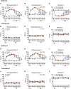
FIGURE 1
FIGURE 1
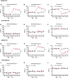
Effects of DI on CFA-induced mice inflammatory pain model. (A,B) The effect on mechanical allodynia (0.07 or 0.4 g) of the ipsilateral hind paw in male mice. Fgroup (36, 200) = 9.377 for (A), Fgroup (36, 200) = 9.944 for (B). (C) The effect on thermal hyperalgesia ipsilateral hind paw in male mice. Fgroup (36, 200) = 13.93. (D–F) The effect of mechanical allodynia and hyperalgesia of the contralateral hind paw. Fgroup (36, 200) = 0.3595 for (D), Fgroup (36, 200) = 0.3423 for (E), Fgroup (36, 200) = 0.5646 for (F). (G–H) The effect on mechanical allodynia (0.07 or 0.4 g) of the ipsilateral hind paw in female mice. Fgroup (36, 200) = 5.347 for (G), Fgroup (36, 200) = 4.478 for (H). (I) The effect on thermal hyperalgesia ipsilateral hind paw in female mice. Fgroup (36, 200) = 6.262. (J–L) The effect of mechanical allodynia and hyperalgesia of the contralateral hind paw. Fgroup (36, 200) = 0.4229 for (J), Fgroup (36, 200) = 0.9263 for (K), Fgroup (36, 200) = 1.391 for (L). Data values are expressed as mean ± SEM. N = 6 mice/group. ** p < 0.01 versus the vehicle group; # p < 0.05, ## p < 0.01 versus the CFA group; ˄p < 0.05, ˄˄p < 0.01 versus the CFA + PBS group.
FIGURE 2
FIGURE 2
Effects of DI pretreatment on CFA-induced mice inflammatory pain model. (A,B) The effect on mechanical allodynia (0.07 or 0.4 g) of the ipsilateral hind paw in male mice. Fgroup (14, 80) = 26.60 for (A), Fgroup (14, 80) = 15.17 for (B). (C) The effect on thermal hyperalgesia ipsilateral hind paw in male mice. Fgroup (14, 80) = 24.28 for (C). (D–F) The effect of mechanical allodynia and hyperalgesia of the contralateral hind paw. Fgroup (14, 80) = 0.8967 for (D), Fgroup (14, 80) = 0.8594 for (E), Fgroup (14, 80) = 1.560 for (F). (G,H) The effect on mechanical allodynia (0.07 or 0.4 g) of the ipsilateral hind paw in female mice. Fgroup (14, 80) = 16.72 for (G), Fgroup (14, 80) = 12.55 for (H). (I) The effect on thermal hyperalgesia ipsilateral hind paw in female mice. Fgroup (14, 80) = 17.73 for (I). (J–L) The effect of mechanical allodynia and hyperalgesia of the contralateral hind paw. Fgroup (14, 80) = 0.9991 for (J), Fgroup (14, 80) = 1.161 for (K), Fgroup (14, 80) = 1.198 for (L). Data values are expressed as mean ± SEM. N = 6 mice/group. **p < 0.01 versus the vehicle group.
FIGURE 3
FIGURE 3
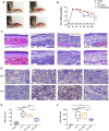
Effect of DI on paw thickness and Inflammatory cell infiltration in CFA model. (A) Macroscopic images of hind paw derived from vehicle, CFA and CFA + PBS and CFA + DI-treated mice. (B) Hind paw thickness (mm) as a function of time (days) was measured in each group. Fgroup (18, 105) = 22.88 for (B) (N = 6 mice/group). (C) HE staining. Inflammation with abundant lymphocytes and sparse neutrophilic granulocytes in a CFA mouse with hypodermic injection and low-grade inflammation in a CFA mouse with a hypodermic injection of DI (20 mg). This experiment was repeated independently 3 times and similar results were obtained. The scale bar represents 200 and 100 μm. (D) Photomicrographs representing MPO and CD45 immunoreactivity in each group. Arrowheads indicate positive cells. ** p < 0.01 versus the vehicle group; # p < 0.05 versus the CFA group; ˄p < 0.05, ˄˄p < 0.01 versus the CFA + PBS group. (E,F) MPO score and CD45 score. Fgroup (3, 16) = 16.68 for (E), Fgroup (3, 16) = 28.36 for (F). The scale bar represents 50 μm. This experiment was repeated independently 3–5 times and similar results were obtained. * p < 0.05, ** p < 0.01 and *** p < 0.001.
FIGURE 4
FIGURE 4
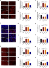
Effect of DI on macrophage and microglia activation. (A) Immunostaining of F4-80 (red) showing macrophages activation in paw tissue. The scale bar represents 100 μm (left) and 50 μm (right). (B) Representative immunofluorescence staining level for F4-80, fold change versus the vehicle group. Fgroup (3, 8) = 16.65 for (B). Real-time PCR were used to detect pro-inflammatory factors IL-1β, Fgroup (3, 12) = 11.87 for (C), TNF-α, Fgroup (3, 8) = 8.567 for (D) and anti-inflammatory factor IL-10, Fgroup (3, 8) = 18.75 for (E). (F) Immunostaining of F4-80 (red) and DAPI (blue) showing macrophages activation in DRG. The scale bar represents 100 μm (left) and 50 μm (right). (G) Representative immunofluorescence staining level for F4-80, fold change versus the vehicle group. Fgroup (3, 12) = 18.83 for (G) .Real-time PCR were used to detect pro-inflammatory factors IL-1β, Fgroup (3, 12) = 20.84 for (H), TNF-α, Fgroup (3, 12) = 11.49 for (I) and anti-inflammatory factor IL-10, Fgroup (3, 12) = 6.271 for (J). (K) Immunostaining of IBA-1 (red) showing microglia activation in spinal cord. Fgroup (3, 8) = 19.96 for (K). The scale bar represents 200 μm (left) and 100 μm (right). (L) Representative immunofluorescence staining level for IBA-1, fold change versus the vehicle group. Real-time PCR analysis of IL-1β, Fgroup (3, 8) = 10.92 for (M), TNF-α, Fgroup (3, 8) = 8.719 for (N) and IL-10, Fgroup (3, 12) = 23.67 for (O) expression relative to vehicle levels in spinal cord. This experiment was repeated independently 3 times and similar results were obtained. * p < 0.05, ** p < 0.01 and *** p < 0.001.
FIGURE 5
FIGURE 5
Effect of DI on macrophage/microglia polarization. (A) Photomicrographs representing iNOS and Arg1 immunoreactivity in each group. Arrowheads indicate positive cells. (B) iNOS score and Arg1 score were detected the same as above. Fgroup (3, 12) = 113.0 for iNOS. Fgroup (3, 12) = 42.18 for Arg1. The scale bar represents 50 μm. (C,E) Immunofluorescent staining for CD68 (Green), CD86 (red), and CD206 (red), and nuclei were counterstained using DAPI (blue). The scale bar represents 50 μm. Representative immunofluorescence staining level for CD86+/CD68+, Fgroup (3, 8) = 28.14 for (D) and CD206+/CD68+, Fgroup (3, 8) = 9.312 for (F), fold change versus the vehicle group. Immunostaining of CD86 (G) and CD206 (I) in DRG. The scale bar represents 50 μm. Representative immunofluorescence staining level for CD86, Fgroup (3, 8) = 11.12 for (H) and CD206, Fgroup (3, 8) = 8.999 for (J), fold change versus the vehicle group. (K) Immunostaining of CD206 in the spinal cord. The scale bar represents 50 μm. (L) Representative immunofluorescence staining level for CD206, Fgroup (3, 8) = 12.42, fold change versus the vehicle group. This experiment was repeated independently 3 times and similar results were obtained. * p < 0.05, ** p < 0.01 and *** p < 0.001.
FIGURE 6
FIGURE 6
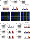
DI inhibited NLRP3 inflammasome activation and IL-1β secretion. (A,B) Western blotting bands and analysis of NLRP3, ASC, NF-κB in plantar tissue. Fgroup (3, 8) = 10.95 for NLRP3, Fgroup (3, 8) = 18.02 for ASC, Fgroup (3, 8) = 12.64 for NF-κB. (C,D) Western blotting bands and analysis of Caspase1 p45 and p20 in plantar tissue. Fgroup (3, 12) = 9.598 for Caspase1 p45, Fgroup (3, 12) = 16.47 for Caspase1 p20. (E,F) Western blotting bands and analysis of pro-IL-1β and IL-1β p17 in plantar tissue. Fgroup (3, 20) = 11.32 for pro-IL-1β, Fgroup (3, 20) = 7.736 for IL-1β p17. Immunofluorescent staining for ASC (green) (G), Caspase-1 (green) (H), IL-1β (green) (I), the nuclei were stained blue with DAPI. The scale bar indicates 50 μm. (J,K) Western blotting bands and analysis of NLRP3, ASC, NF-κB in DRG. Fgroup (3, 16) = 11.44 for NLRP3, Fgroup (3, 16) = 23.45 for ASC, Fgroup (3, 16) = 29.99 for NF-κB. (L,M) Western blotting bands and analysis of Caspase1 p45 and p20 in DRG. Fgroup (3, 16) = 8.323 for Caspase1 p45, Fgroup (3, 16) = 12.98 for Caspase1 p20. (N,O) Western blotting bands and analysis of pro- IL-1β and IL-1β p17 in DRG. Fgroup (3, 16) = 9.598 for pro-IL-1β, Fgroup (3, 12) = 7.633 for IL-1β p17. (P,Q) Western blotting bands and analysis of NLRP3, ASC, NF-κB in spinal cord. Fgroup (3, 16) = 9.572 for NLRP3, Fgroup (3, 16) = 10.55 for ASC, Fgroup (3, 16) = 10.52 for NF-κB. (R,S) Western blotting bands and analysis of Caspase1 p45 and p20 in spinal cord. Fgroup (3, 12) = 17.36 for Caspase1 p45, Fgroup (3, 12) = 13.11 for Caspase1 p20. (T,U) Western blotting bands and analysis of pro- IL-1β and IL-1β p17 in spinal cord. Fgroup (3, 16) = 21.28 for pro-IL-1β, Fgroup (3, 16) = 10.23 for IL-1β p17. N = 3-5 biological repeats, 2 mice/group/repeat. * p < 0.05, ** p < 0.01 and *** p < 0.001.
FIGURE 7
FIGURE 7
(A)The enhanced microglia proliferation in the spinal cord and the infiltration of macrophages into the DRG and paw tissue of the CFA mice contribute to the development of inflammatory pain. (B) DI regulated and inhibited the imbalance between pro-inflammatory and anti-inflammatory responses. (C) DI suppressed the assembly of NLRP3 inflammasome complexes and NF-κB, thereby inhibiting the secretion of IL-1β.

Similar articles

Cited by

References

    1. Allen B. L., Montague-Cardoso K., Simeoli R., Colas R. A., Oggero S., Vilar B., et al. (2020). Imbalance of Pro-resolving Lipid Mediators in Persistent Allodynia Dissociated from Signs of Clinical Arthritis. Pain 161 (9), 2155–2166. 10.1097/j.pain.0000000000001908 - DOI - PMC - PubMed
    1. Bambouskova M., Gorvel L., Lampropoulou V., Sergushichev A., Loginicheva E., Johnson K., et al. (2018). Electrophilic Properties of Itaconate and Derivatives Regulate the IκBζ-ATF3 Inflammatory axis. Nature 556 (7702), 501–504. 10.1038/s41586-018-0052-z - DOI - PMC - PubMed
    1. Bannister K., Kucharczyk M., Dickenson A. H. (2017). Hopes for the Future of Pain Control. Pain Ther. 6 (2), 117–128. 10.1007/s40122-017-0073-6 - DOI - PMC - PubMed
    1. Binshtok A. M., Wang H., Zimmermann K., Amaya F., Vardeh D., Shi L., et al. (2008). Nociceptors Are Interleukin-1beta Sensors. J. Neurosci. 28 (52), 14062–14073. 10.1523/JNEUROSCI.3795-08.2008 - DOI - PMC - PubMed
    1. Chan A. H., Schroder K. (2020). Inflammasome Signaling and Regulation of Interleukin-1 Family Cytokines. J. Exp. Med. 217 (1). 10.1084/jem.20190314 - DOI - PMC - PubMed

LinkOut - more resources