. 2022 Jul 22:13:898067.
doi: 10.3389/fphar.2022.898067. eCollection 2022.

Peripheral Neuroprotective and Immunomodulatory Effects of 5α-Reductase Inhibitors in Parkinson's Disease Models

Affiliations

Peripheral Neuroprotective and Immunomodulatory Effects of 5α-Reductase Inhibitors in Parkinson's Disease Models

Andrée-Anne Poirier et al. Front Pharmacol. .

Abstract

Gastrointestinal disorders in Parkinson's disease (PD) have been associated with neuronal alteration in the plexus of the gut. We previously demonstrated the immunomodulatory effect of female hormones to treat enteric neurodegeneration in the 1-methyl-4-phenyl-1,2,3,6-tetrahydropyridine (MPTP) mouse model of PD. This study made the hypothesis of obtaining similar neuroprotection as with hormone treatments by affecting steroidogenesis with two 5α-reductase inhibitors, finasteride and dutasteride. These drugs are approved to treat benign prostatic hyperplasia and alopecia and display mitochondrial effects. In MPTP-treated mice, the dopaminergic and vasoactive intestinal peptide (VIP) neurons alteration was prevented by finasteride and dutasteride, while the increase in proinflammatory macrophages density was inhibited by dutasteride treatment but not finasteride. NF-κB response, oxidative stress, and nitric oxide and proinflammatory cytokines production in vitro were only prevented by dutasteride. In addition, mitochondrial production of free radicals, membrane depolarization, decreased basal respiration, and ATP production were inhibited by dutasteride, while finasteride had no effect. In conclusion, the present results indicate that dutasteride treatment prevents enteric neuronal damages in the MPTP mouse model, at least in part through anti-inflammatory and mitochondrial effects. This suggests that drug repurposing of dutasteride might be a promising avenue to treat enteric neuroinflammation in early PD.

Keywords: MPTP; dutasteride; enteric nervous system; female hormones; finasteride; gut; inflammation; mitochondria.


Conflict of interest statement

The authors declare that the research was conducted in the absence of any commercial or financial relationships that could be construed as a potential conflict of interest.

Figures

FIGURE 1
FIGURE 1
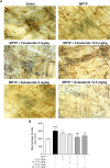
Finasteride and dutasteride treatments prevented MPTP-induced DA neurons damage in the myenteric plexus. (A) Photomicrographs of neuronal ganglia (light blue staining) and TH immunochemistry staining (brown DAB staining) in the myenteric plexus of the distal ileum. The white arrowheads indicate TH+ cell bodies; scale bar = 25 µm. (B) Stereological counts of TH+ neurons in the myenteric plexus showed neuroprotective effect of finasteride (Fin) (12.5 mg/kg) and dutasteride (Dut) (5 mg/kg) in MPTP mice. Values shown are the average densities (per mm2) ± SEM of 10 mice per group. * p < 0.05 and **** p < 0.0001 compared to saline control mice; ## p < 0.01 and #### p < 0.0001 compared to MPTP lesioned mice; ψ p < 0.05 compared to MPTP + finasteride (5 mg/kg) treated mice.
FIGURE 2
FIGURE 2
Protection of VIP neurons in the myenteric plexus of MPTP mice with finasteride and dutasteride. (A) Photomicrographs of VIP immunofluorescence staining in the myenteric plexus of the distal ileum; scale bar = 25 µm. (B) VIP immunoreactivity showed neuroprotective effect of finasteride (Fin) (12.5 mg/kg) and dutasteride (Dut) (5 mg/kg) in MPTP mice. Values shown are the average densities (per mm2) ± SEM of 8–10 mice per group. ** p < 0.01 compared to saline controls mice; # p < 0.05 compared to MPTP lesioned mice; ψ p < 0.05 compared to MPTP + finasteride (5 mg/kg) treated mice.
FIGURE 3
FIGURE 3
Dutasteride treatment prevented the MPTP-induced macrophage response in the myenteric plexus, whereas finasteride gave an inconclusive effect. (A) Photomicrographs of Iba-1 immunochemistry staining (brown DAB staining) in the myenteric plexus of the distal ileum; scale bar = 25 µm. (B) Stereological counts of macrophages in the myenteric plexus showed an MPTP-induced increase in macrophage density inhibited by dutasteride (Dut) treatment (5 and 12.5 mg/kg), whereas finasteride (Fin) treatment at 5 mg/kg was ineffective and at 12.5 mg/kg led to no change compared to controls or vehicle-treated MPTP mice. Values shown are the average densities (per mm2) ± SEM of 10 mice per group. * p < 0.05 and **** p < 0.0001 compared to saline control mice; # p < 0.05 and ## p < 0.01 compared to MPTP lesioned mice.
FIGURE 4
FIGURE 4
Proinflammatory macrophages density increased by MPTP was reduced by dutasteride treatment in the myenteric plexus, whereas finasteride gave an inconclusive effect. (A) Photomicrographs of MHCII immunofluorescence labeling in the myenteric plexus of the distal ileum; scale bar = 25 µm. (B) Stereological counts of MHCII+ cells showed that dutasteride (Dut) (5 and 12.5 mg/kg) prevented the increase of proinflammatory macrophages in the myenteric plexus of MPTP mice, whereas finasteride treatment at 5 mg/kg was ineffective and at 12.5 mg/kg led to no change compared to controls or vehicle-treated MPTP mice. Values shown are the average densities (per mm2) ± SEM of 10 mice per group. **** p < 0.0001 compared to saline control mice; ## p < 0.01 and ### p < 0.001 compared to MPTP lesioned mice; ψψ p < 0.01 and ψψψψ p < 0.0001 compared to MPTP + finasteride (5 mg/kg) treated mice.
FIGURE 5
FIGURE 5
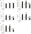
Dutasteride but not finasteride treatment inhibited the induction of NF-κB proinflammatory response by MPP+ in a human monocytic cell line. NF-κB activity was determined in the supernatant of monocytic cells treated with finasteride (Fin) or dutasteride (Dut) combined or not with the MPP+ toxin. Using THP1-XBlue cells containing NF-κB reporter gene allowed the evaluation of NF-κB activation level in this human cell line. Histogram show (A) THP1-XBlue cell viability quantification (MTT assay) and (B) NF-κB response as percentage of negative control. Results are the mean of three experiments. * p < 0.05, ** p < 0.01, *** p < 0.001 compared to control; ## p < 0.01 and #### p < 0.0001 compared to MPP+ treatment; ψ p < 0.05 and ψψ p < 0.01 compared to finasteride only.
FIGURE 6
FIGURE 6
Significant reduction of proinflammatory markers by dutasteride but not by finasteride treatment in THP-1 cells following MPP+ stimulation. Nitric oxide (NO), IL-1β, and IL-6 production was measured in the supernatant of THP-1 monocytic cells treated with finasteride (Fin) or dutasteride (Dut) in the presence or the absence of MPP+. Histograms show (A) THP-1 cell viability quantification (MTT assay) and (B) NO accumulation by THP-1 cells (Griess assay) as percentage of negative control, (C) IL-1β and (D) IL-6 cytokine production in pg/ml, and (E) number of positive THP-1 cells to the oxidative stress marker CellROX Orange as percentage of cells. Results are the mean of three experiments. * p < 0.05 and **** p < 0.0001 compared to control; #### p < 0.0001 compared to MPP+ treatment; ψψ p < 0.01 and ψψψψ p < 0.0001 compared to Finasteride only; ξξξξ p < 0.0001 compared to MPP+ + finasteride.
FIGURE 7
FIGURE 7
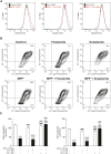
Impaired mitochondrial function was prevented by dutasteride but not by finasteride treatment in MPP+-stimulated THP-1 cells. Three parameters of mitochondrial function were evaluated with finasteride- (Fin) or dutasteride-treated (Dut) THP-1 cells exposed or not to the MPP+ toxin. Flow cytometry analyses were used to measure mitochondrial oxidative stress and membrane potential, while oxygen consumption rate (OCR) was quantified by Seahorse assays. (A) An increase in the mean fluorescence intensity (MFI; values in parentheses) of the marker MitoSOX red reagent corresponds to an increase in mitochondrial oxidative stress production (shift of the curve to the right). (B) The JC-1 dye enables evaluation of the mitochondrial membrane potential; JC-1 green intensity increases with membrane depolarization, while JC-1 red intensity increases with polarized membranes. (C) Histograms show mitochondrial basal respiration and ATP production quantification in each condition (pmol/min). Results are the mean of three experiments. **** p < 0.0001 compared to control; #### p < 0.0001 compared to MPP+ treatment; ψ p < 0.05, ψψ p < 0.01 and ψψψψ p < 0.0001 compared to finasteride only; ξξξξ p < 0.0001 compared to MPP+ + finasteride.
FIGURE 8
FIGURE 8
Summary of the effects of finasteride and dutasteride in the enteric nervous system of the MPTP mouse model of Parkinson’s disease. Schematic interpretation of the effect of finasteride (Fin) or dutasteride (Dut) treatments in the myenteric plexus of the MPTP mouse model according to the present results (illustration adapted from Côté et al., 2011). Upper panel illustrates the MPTP-induced damages in the myenteric plexus (previously investigated in (25)). (A) MPTP enters myenteric ganglia and is processed by enteroglial cells into MPP+, (B) the latter is recaptured by macrophages and leads to Iba-1+MHCII+ proinflammatory cell polarization, or by TH+ and VIP+ neurons and leads to cell damage (C). This neuronal damage induces an immune response from Iba-1+ resident macrophages and the release of chemokines (D). The resulting chemokine gradient promotes the detrimental infiltration of monocytes (E). These events are associated with monocyte differentiation (F) into proinflammatory macrophages (Iba-1+MHCII+) that may subsequently induce neuronal damage (G). Lower panels are close-ups of the individual effects of finasteride or dutasteride in each cell type studied. (1) MPTP-induced TH+ neuron alteration is completely inhibited by finasteride and dutasteride treatments. (2) Finasteride and dutasteride inhibit VIP+ neuron damage. Dutasteride administration inhibits both Iba-1+ resident macrophage recruitment (3) and Iba-1+MHCII+ proinflammatory macrophage polarization (4) following MPTP injections, while finasteride had no effect. (5) Finally, dutasteride treatment completely inhibits activation of the NF-κB transcription factor, cellular production of oxidative stress (ROS), production of the proinflammatory cytokines IL-1β and IL-6, mitochondrial membrane depolarization, and mitochondrial free radical production (ROS) in monocytes. The arrows indicate the consequences of administering MPTP, while the closed ended lines show an inhibition of the effect.

Similar articles

Cited by

References

    1. Annerino D. M., Arshad S., Taylor G. M., Adler C. H., Beach T. G., Greene J. G. (2012). Parkinson's Disease Is Not Associated with Gastrointestinal Myenteric Ganglion Neuron Loss. Acta Neuropathol. 124, 665–680. 10.1007/s00401-012-1040-2 - DOI - PMC - PubMed
    1. Azzolina B., Ellsworth K., Andersson S., Geissler W., Bull H. G., Harris G. S. (1997). Inhibition of Rat Alpha-Reductases by Finasteride: Evidence for Isozyme Differences in the Mechanism of Inhibition. J. Steroid Biochem. Mol. Biol. 61, 55–64. 10.1016/s0960-0760(97)00002-2 - DOI - PubMed
    1. Azzouni F., Godoy A., Li Y., Mohler J. (2012). The 5 Alpha-Reductase Isozyme Family: a Review of Basic Biology and Their Role in Human Diseases. Adv. Urol. 2012, 530121. 10.1155/2012/530121 - DOI - PMC - PubMed
    1. Badanjak K., Fixemer S., Smajić S., Skupin A., Grünewald A. (2021). The Contribution of Microglia to Neuroinflammation in Parkinson's Disease. Int. J. Mol. Sci. 22, 4676. 10.3390/ijms22094676 - DOI - PMC - PubMed
    1. Borsche M., König I. R., Delcambre S., Petrucci S., Balck A., Brüggemann N., et al. (2020). Mitochondrial Damage-Associated Inflammation Highlights Biomarkers in PRKN/PINK1 Parkinsonism. Brain 143, 3041–3051. 10.1093/brain/awaa246 - DOI - PMC - PubMed

LinkOut - more resources