. 2022 Jul 22:13:692437.
doi: 10.3389/fphar.2022.692437. eCollection 2022.

Effect of Acid Suppressants on Non- Helicobacter pylori Helicobacters Within Parietal Cells

Affiliations

Effect of Acid Suppressants on Non- Helicobacter pylori Helicobacters Within Parietal Cells

Masahiko Nakamura et al. Front Pharmacol. .

Abstract

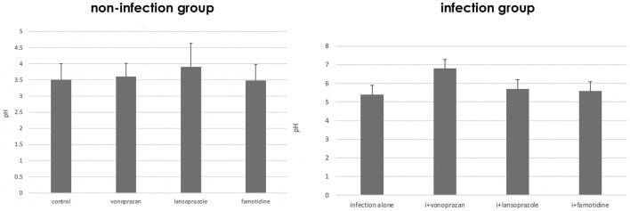
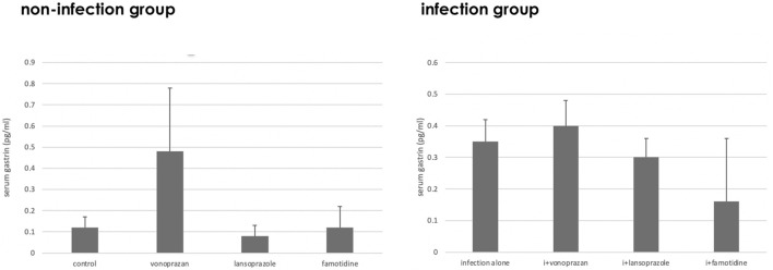
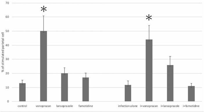
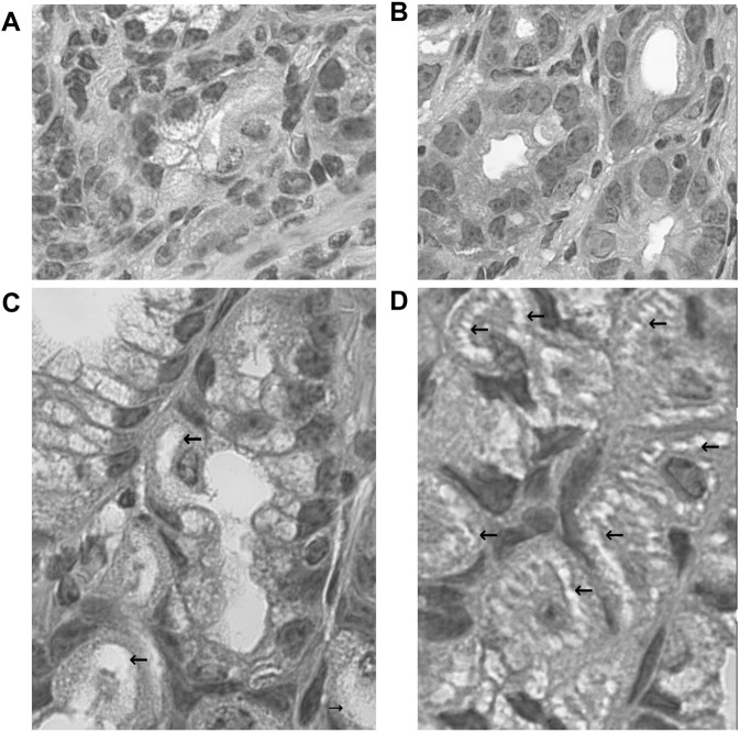
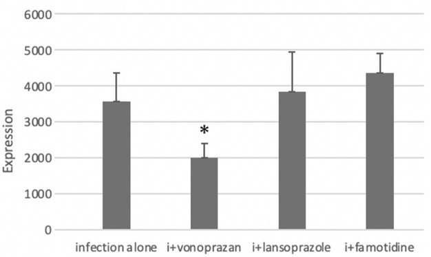
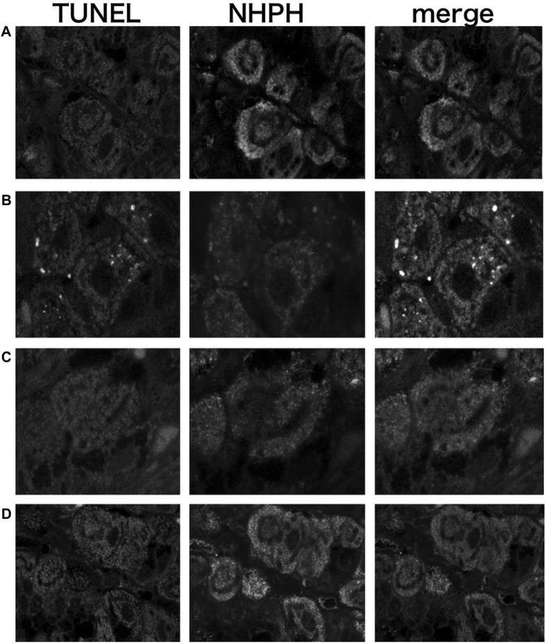
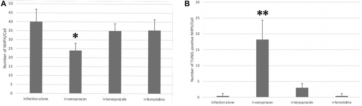
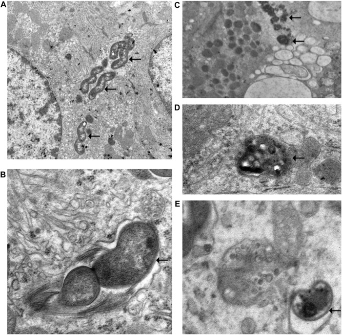
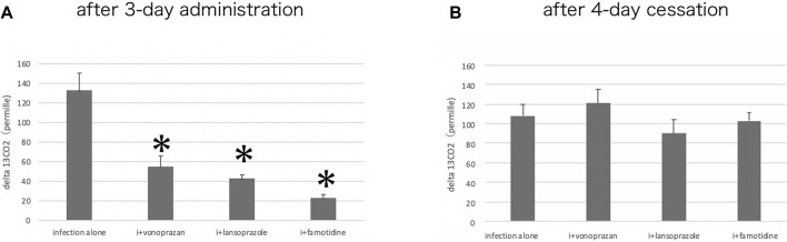
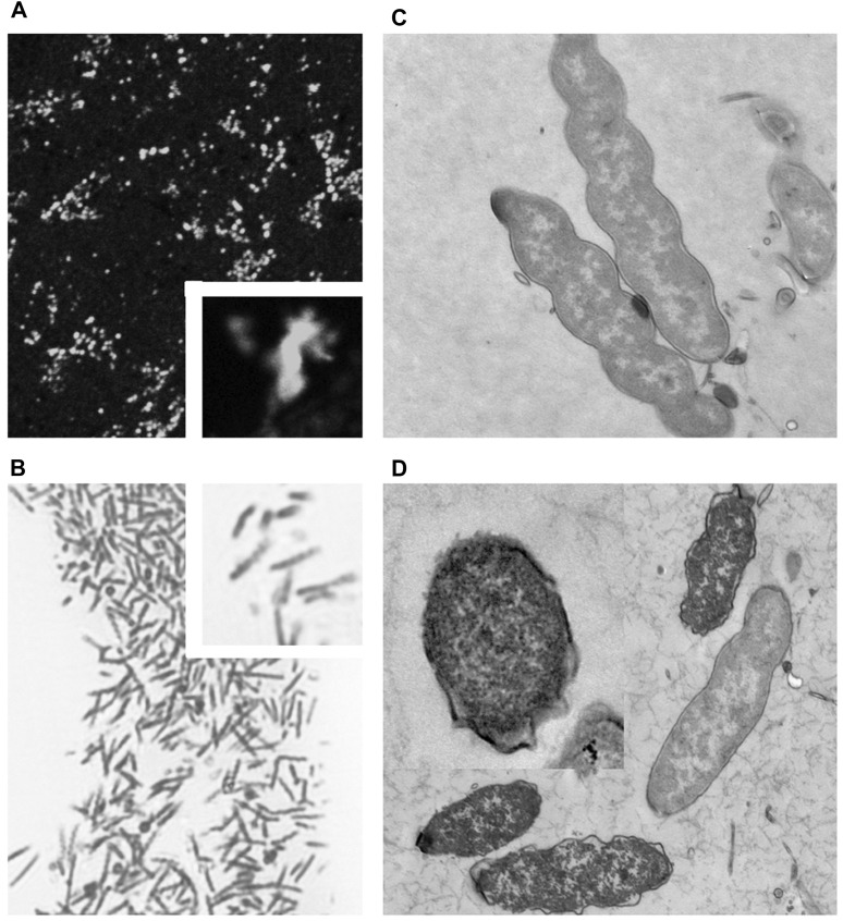
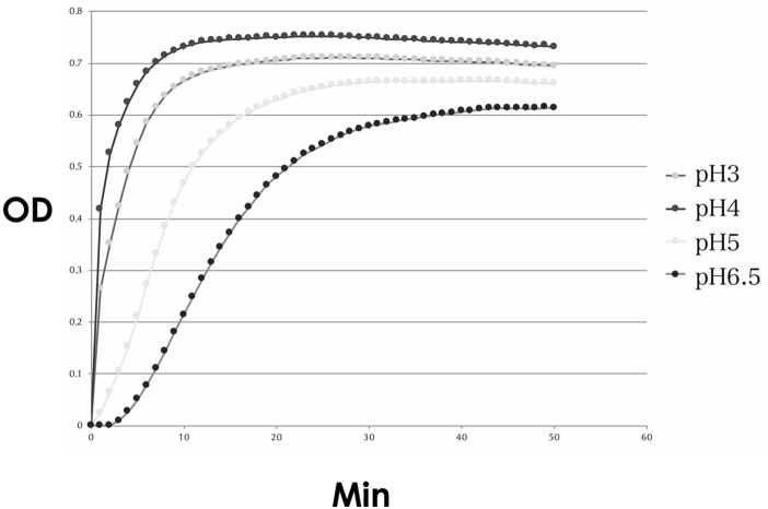
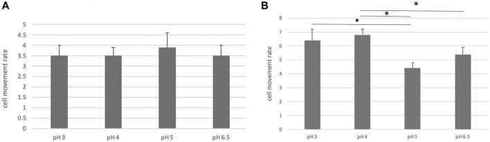
We investigated the effect of increased pH induced by acid suppressants on the viability of non-Helicobacter pylori helicobacters (NHPHs) within parietal cell intracellular canaliculi and fundic glandular lumina by immunohistochemistry, electron microscopy, quantitative PCR, urea breath tests, and using a bilayer culture system. Three months before the experiment, mice were infected with the NHPH H. suis and then treated with famotidine (2 mg/kg body weight [BW], once daily), lansoprazole (30 mg/kg BW, once daily), or vonoprazan (20 mg/kg BW, once daily) for 3 days. Immunohistochemical studies using the TUNEL method, quantitative PCR analysis, and urea breath tests were performed. PCR analysis showed a decrease in the NHPH quantity after vonoprazan treatment. Urea breath tests revealed a significant decrease in the NHPH urease activity after vonoprazan, lansoprazole, and famotidine treatments for 3 days; however, 4 days after the treatment, urease activity reversed to the pretreatment level for each treatment group. Electron microscopy revealed an increase in the damaged NHPH after vonoprazan treatment. The TUNEL method revealed apoptotic NHPH within parietal cells after vonoprazan treatment. The bilayer culture results demonstrated that NHPH moved more quickly at a pH of 4.0 than at a pH of 3.0, 5.0, and 6.5, and electron microscopy revealed a change from the spiral form to the coccoid form under near-neutral pH conditions. We thus proposed that acid suppressants, especially vonoprazan, induce NHPH damage by altering pH.

Keywords: Helicobacter suis; TUNEL; bilayer culture; coccoid form; non-Helicobacter pylori helicobacter; urea breath test; vonoprazan.


Conflict of interest statement

The authors declare that the research was conducted in the absence of any commercial or financial relationships that could be construed as a potential conflict of interest.

Figures

FIGURE 1
FIGURE 1
Alteration of gastric pH by acid suppressants and NHPH infection. A significant interaction was found between infection and acid suppressants in the gastric mucosal pH (F = 3.04 and p = 0.043 by two-way ANOVA). In the infection group, vonoprazan treatment induced the highest increase in intragastric pH. i means infection in the left graph.
FIGURE 2
FIGURE 2
Alteration of the serum gastrin level by acid suppressants and NHPH infection. A significant interaction was found between infection and acid suppressants in the serum gastrin level (F = 5.61; p = 0.003).
FIGURE 3
FIGURE 3
Alteration of the parietal cell structure by acid suppressants and NHPH infection. No significant interaction was found between infection and acid suppressants in the percentage of the stimulated parietal cell (F = 1.54; p = 0.224). By the post hoc analysis, the vonoprazan-treated group showed a significant increase in the stimulated parietal cell in both non-infection and infection groups (p < 0.05).
FIGURE 4
FIGURE 4
Morphological changes of fundic glandular cells by acid suppressants under NHPH infection. HE stains (A) the control group fundic mucosa, X 400. (B) In famotidine-treated mice, no significant changes in the parietal cell were detected in the body portion of the fundic gland, X 400. (C) In the lansoprazole-treated mice, many parietal cells having very enlarged intracellular canaliculi (arrows) were seen, X 600. (D) In the vonoprazan-treated mice, most parietal cells had the enlarged intracellular canaliculi (arrows) surrounding the nucleus, X 600.
FIGURE 5
FIGURE 5
Alteration of NHPH localization in the fundic gland in the infection group by acid suppressants by indirect fluorescent immunohistochemistry using an anti-HsvA antibody. (A) In the infection-alone group, linear-shaped NHPHs were mostly found within the intracellular canaliculi of the parietal cells and in the gastric glandular lumen, X 400. (B) In the famotidine-treated mice, many spiral-shaped NHPHs were seen in the intracellular canaliculi of the parietal cells and in the gastric glandular lumen, X 400. (C) In the lansoprazole-treated mice, many spiral-shaped NHPHs were seen in the enlarged intracellular canaliculus of the parietal cells and in the gastric glandular lumen, X 600. (D) In the vonoprazan-treated mice, the round-shaped NHPHs were mostly seen in the intracellular canaliculus of the parietal cells, X 400.
FIGURE 6
FIGURE 6
Effect of acid suppressants on the NHPH number by PCR. PCR analysis showed significant difference between infection alone and administered agents (F = 5.33 and p = 0.008 by one-way ANOVA). Post hoc analyses (Bonferroni correction) revealed that the infection-alone group is significantly larger than the infection + vonoprazan group (*p = 0.008). Decrease in NHPH in the vonoprazan-treated mice, but significant interaction was found between infection and administered agents in NHPHs (F = 7.17; p = 0.008).
FIGURE 7
FIGURE 7
Localization of apoptotic NHPHs in the infection group by acid suppressants by the TUNEL method X 200. (A) In the infection-alone group, no apoptosis was detected. (B) In the vonoprazan-treated group, apoptotic NHPHs were detected within the intracellular canaliculi of the parietal cells. (C) In the lansoprazole-treated group, many NHPHs were found, but no apoptotic reaction was recognized. (D) In the famotidine-treated group, NHPHs in the parietal cells were negative for the TUNEL reaction.
FIGURE 8
FIGURE 8
Quantitative analysis of all NHPHs and apoptotic NHPHs by using acid suppressants by immunohistochemistry. (A) Significant difference was found between infection alone and administered agents in the NHPH number (F = 6.14; p = 0.005 by one-way ANOVA). Post hoc analyses (Bonferroni correction) revealed that the infection-alone group is significantly larger than the infection + vonoprazan group (*p = 0.004). (B) Significant difference was found between infection alone and administered agents in the TUNEL-positive NHPH number (F = 28.1; p < 0.001 by one-way ANOVA). Post hoc analyses (Bonferroni correction) revealed that the infection + vonoprazan group is significantly larger than the infection-alone group (**p < 0.001).
FIGURE 9
FIGURE 9
Electron microscopic observation of NHPH in infection-alone and infection + vonoprazan-treated mouse gastric parietal cells. (A) In infection alone mouse, many NHPHs were seen within the intracellular canaliculus of the parietal cell in the gastric mucosa, X 8,000. (B) In a higher magnification, flagella were recognized adjacent to the NHPH cell body in the intracellular canaliculus, X 24,000. (C) In the infection + vonoprazan-treated mouse, the bacterial cell body became condensed and irregular (arrows) in the intracellular canaliculus of the parietal cell, X 8,000. (D,E) Many bacilli were found in the secondary lysosome in the infection + vonoprazan-treated group, X 8,000.
FIGURE 10
FIGURE 10
Alteration of the urease activity by acid suppressants. (A) By the urea breath test, a significant interaction was found between infection alone and administered agents (F = 47.1; p = 0.001 by one-way ANOVA). Post hoc analyses (Bonferroni correction) revealed that the infection-alone group is significantly larger than the other three groups (*p < 0.001) after three-day administration. (B) Cessation of the administration for 4 days resulted in the recovery of the urease activity in all drug-administered groups.
FIGURE 11
FIGURE 11
Fluorescent, light, and electron microscopic observations of the cultured NHPH (A) By fluorescent immunohistochemical observation of the cultured NHPH using the HsvA antibody, many bacteria were observed in the incubation medium, X 400. Inset X 1800. (B) By light microscopic observation of the toluidine-blue stained Epon-embedded 2-µM section, many spiral bacilli were observed, X 600. Inset X 1800. (C) Electron microscopic observation of the cultured intact NHPH in pH 3. Intact bacilli were observed with flagella, X 15, 000. Ruthenium red en bloc staining. (D) Electron microscopic observation of the cultured NHPH in pH 6.5. Ruthenium red en bloc staining. The cytoplasm of some of the bacilli became condensed and homogeneous, and its plasma membrane became irregular, coinciding with the observation reported in the coccoid form, X 12,000, Inset x15, 000.
FIGURE 12
FIGURE 12
Urease activity of the cultured NHPH. Urease activity was found to be strongest at pH 4, followed by pH 3, and became weak at pH 5 and 6.5. OD: optical density.
FIGURE 13
FIGURE 13
Motility of the cultured NHPH. On day 2, no significant difference was found, as shown in (A), while a significant decrease in motility was detected at pH 5 and 6.5 on day 3 (B) by post hoc analysis (*<0.05).

Similar articles

Cited by

References

    1. Azevedo N.F., Almeida C., Cerqueira L., Dias S., Keevil C.W., Vieira M.J. (2007). Coccoid form of Helicobacter pylori as a morphological manifestation of cell adaptation to the environment. Appl Environ Microbiol 73, 3423–3427. Epub 2007 Mar 30. 10.1128/AEM.00047-07 - DOI - PMC - PubMed
    1. Bell NJ, Hunt RH. (1996). Time to maximum effect of lansoprazole on gastric pH in normal male volunteers. Aliment Pharmacol Ther 10, 897–904. PMID: 8971286. 10.1046/j.1365-2036.1996.103242000.x - DOI - PubMed
    1. Bizzozero G. (1893). Ueber die schlauchförmigen Drüsen des Magendarmkanals und die Beziehungen ihres Epithels zu dem Oberflächenepithel der Schleimhaut Dritte Mittheilung. Archiv f. mikrosk. Anat. 42, 82–152. 10.1007/bf02975307 - DOI
    1. Calhoun BC, Goldenring JR. (1996). Rab proteins in gastric parietal cells: evidence for the membrane recycling hypothesis. Yale J Biol Med 69, 1–8. PMID: 9041684; PMCID: PMC2588980. - PMC - PubMed
    1. Chu S, Tanaka S, Kaunitz JD, Montrose MH. (1999). Dynamic regulation of gastric surface pH by luminal pH. J Clin Invest 103, 605–12. 10.1172/JCI5217 - DOI - PMC - PubMed

LinkOut - more resources