. 2021 Jun:46:1-25.
doi: 10.3767/persoonia.2021.46.01. Epub 2020 Sep 21.

Diversity of Backusella (Mucoromycotina) in south-eastern Australia revealed through polyphasic taxonomy

Affiliations

Diversity of Backusella (Mucoromycotina) in south-eastern Australia revealed through polyphasic taxonomy

A S Urquhart et al. Persoonia. 2021 Jun.

Abstract

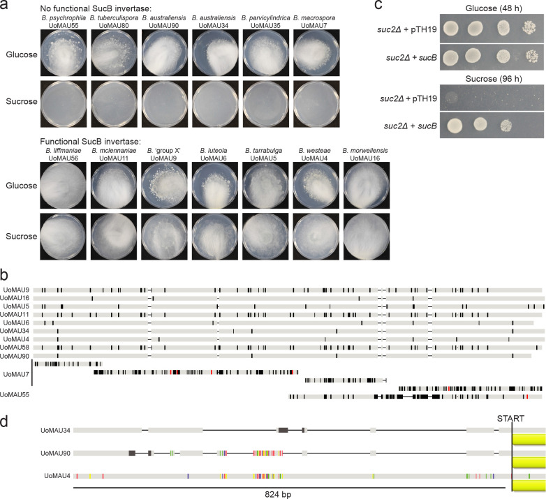
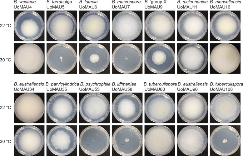
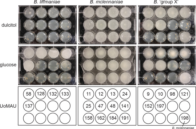
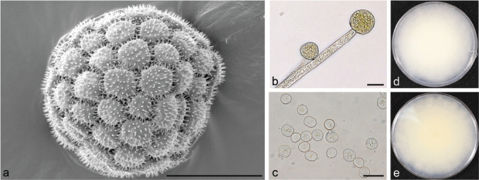
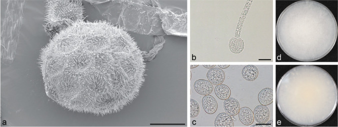
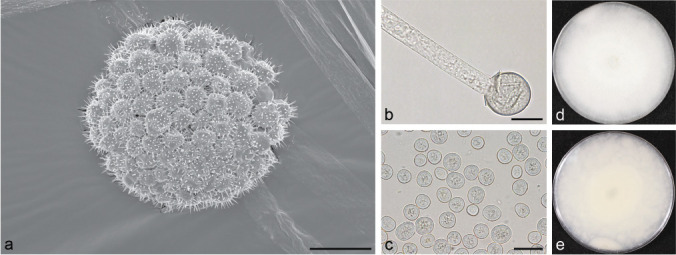
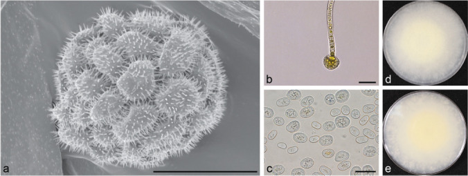
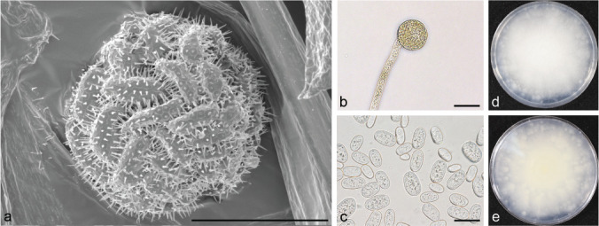
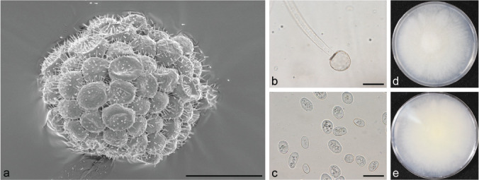
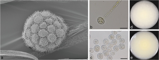
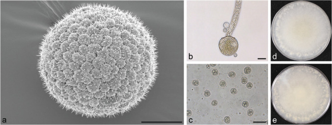
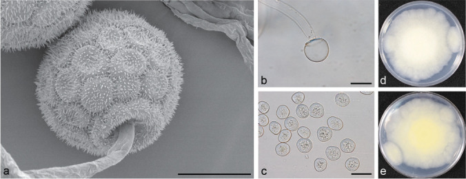
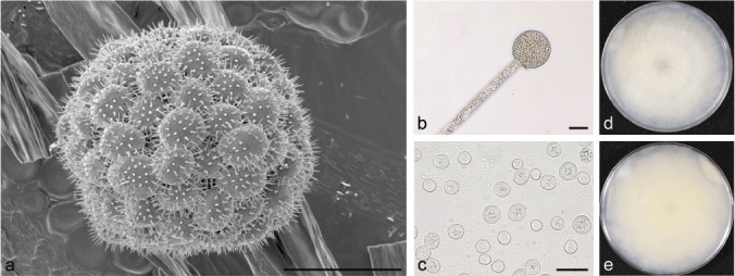
Here we explore the diversity of one morphologically distinguishable genus in the Mucoromycotina, Backusella, in south-eastern Australia. We isolated more than 200 strains from locations across the states of Victoria and Tasmania. Characterization of these strains using a combination of approaches including morphology, sucrose utilization and whole genome sequencing for 13 strains, revealed 10 new species. The genetic basis for interspecies variation in sucrose utilization was found to be the presence of a gene encoding an invertase enzyme. The genus Backusella is revised and a new key for species identification produced. Given that we have more than doubled the number of species in this genus, this work demonstrates that there may be considerable undiscovered species diversity in the early diverging fungal lineages. Citation: Urquhart AS, Douch JK, Heafield TA, et al. 2021. Diversity of Backusella (Mucoromycotina) in south-eastern Australia revealed through polyphasic taxonomy. Persoonia 46: 1-25. https://doi.org/10.3767/persoonia.2021.46.01.

Keywords: Backusella; Mucorales; genome sequencing; invertase; new taxa; polyphasic taxonomy; zygospore.


Figures

Fig. 1
Fig. 1
Strains of Backusella were isolated across the south-eastern corner of Australia. Collections covered a range of habitats including wet sclerophyll forest in: a. Wilson’s Promontory National Park; b. Pittosporum undulatum dominated warm temperate rainforest in Uralla Nature Reserve; c. Nothofagus cunninghamii dominated cool temperate rainforest in Toolangi State Forest; d. dry Eucalyptus woodland in the Brisbane Ranges National Park; e. damp Eucalyptus forest of Jack Cann Reserve. — f. Distribution of collection sites. Map data: Google, SIO, NOAA, U.S. Navy, NGA, GEBCO.
Fig. 2
Fig. 2
UPGMA tree based on partial LSU sequence. Nodes are labelled with % bootstrap support from 1 000 replicates. The bootstrap support values derived from maximum likelihood analysis are given in parentheses based on 1 000 repeats for clades which were supported. Taxa in bold indicate sequences derived from type specimens. First dot represents growth on sucrose: green = utilizes sucrose, red = does not utilize sucrose. Second dot represents growth at 30 °C after 3 d. Red = no growth, amber = less than 2 cm growth, green = more than 2 cm growth.
Fig. 3
Fig. 3
Bayesian phylogeny based on the arginine succinate lyase (argA) region. Taxa in bold indicate sequences derived from type specimens. Mucor circinelloides (Corrochano et al. 2016) and Rhizopus microsporus (Mondo et al. 2017) sequences were used as outgroups. Posterior probabilities are indicated. Bootstrap support values derived from maximum likelihood analysis are given in parentheses based on 1 000 repeats.
Fig. 4
Fig. 4
Bayesian phylogeny based on the ITS region. The letter c followed by a number indicates a sequence from cloned DNA. Taxa in bold indicate sequences derived from type specimens. Backusella oblongispora was used as the outgroup. Sequences with accession numbers were obtained from GenBank. Posterior probabilities are indicated. Bootstrap support values derived from maximum likelihood analysis are given in parentheses based on 1 000 repeats.
Fig. 5
Fig. 5
A multi-locus Bayesian phylogenetic tree resolves two species groups within the genus Backusella in Australia. Branches are labelled with posterior probabilities (%). Bootstrap support values derived from a maximum likelihood analysis are given in parentheses based on 1 000 repeats.
Fig. 6
Fig. 6
Graph of spore dimensions for the strains of Backusella isolated in this study. Q represents the quotient of average spore length and width.
Fig. 7
Fig. 7
Pigmentation of the whole genome sequenced strains of Backusella after scraping from potato dextrose agar culture. Strains are labelled with UoMAU numbers, with the Clade 1 and Clade 2 species indicated. All strains within each species had consistent colony pigmentation conforming to that of the representative stains shown here.
Fig. 8
Fig. 8
Newly described Backusella species are heterothallic. a. Diagram of the mating type locus of the sequenced strains. The mating type of UoMAU7 was fragmented in the assembly, hence one flank is missing; b. SEM of zygospores for the four species for which mating partners were identified. — Scale bars = 20 μm.
Fig. 9
Fig. 9
Sucrose assimilation in Backusella corresponds to the presence of a functional sucB gene encoding invertase. a. Growth of strains on sucrose as the sole carbon source is variable between species; b. strains that are able to utilize sucrose have alleles of sucB with a full open reading frame. Shown is a translated nucleotide alignment with polymorphisms highlighted in black and stop codons in red in UoMAU7 and UoMAU55; c. the sucB gene from B. westeae confers the ability to utilize sucrose to a Saccharomyces cerevisiae SUC2Δ deletion mutant; d. two B. australiensis strains that are unable to grow on sucrose have a sucB allele. One possible explanation is that deletions in the promoter region are affecting expression of the gene, illustrated by the black lines in UoMAU34 and UoMAU90.
Fig. 10
Fig. 10
Backusella strains have variable temperature dependent growth. Growth of representative strains at 22 °C and 30 °C. Note the wide intraspecific variation between B. tuberculispora UoMAU80 and B. tuberculispora UoMAU108.
Fig. 11
Fig. 11
Growth of Backusella ‘group X’, B. mclennaniae and B. liffmaniae on media containing either dulcitol or glucose as a sole carbon source.
Fig. 12
Fig. 12
Morphology of Backusella australiensis strain UoMAU34. a. SEM of sporangium; b. light microscope image of columella; c. light microscope image of sporangiospores; d, e. obverse and reverse of colony. — Scale bars = 20 μm.
Fig. 13
Fig. 13
Morphology of Backusella dispersa strain UoMAU119. a. SEM of sporangium; b. light microscope image of columella; c. light microscope image of sporangiospores; d, e. obverse and reverse of colony. — Scale bars = 20 μm.
Fig. 14
Fig. 14
Backusella dispersa strains produced abundant giant cells in their substrate mycelia that are distinct from the inflated droplet filled hyphal regions seen in other strains like B. tuberculispora and B. westeae. — Scale bar = 100 μm.
Fig. 15
Fig. 15
Morphology of Backusella liffmaniae strain UoMAU58. a. SEM of sporangium; b. light microscope image of columella; c. light microscope image of sporangiospores; d, e. obverse and reverse of colony. — Scale bars = 20 μm.
Fig. 16
Fig. 16
Morphology of Backusella luteola strain UoMAU6. a. SEM of sporangium; b. light microscope image of columella; c. light microscope image of sporangiospores; d, e. obverse and reverse of colony. — Scale bars = 20 μm.
Fig. 17
Fig. 17
Morphology of Backusella macrospora strain UoMAU7. a. SEM of sporangium; b. light microscope image of columella; c. light microscope image of sporangiospores; d, e. obverse and reverse of colony. — Scale bars = 20 μm.
Fig. 18
Fig. 18
Morphology of Backusella mclennaniae strain UoMAU11. a. SEM of sporangium; b. light microscope image of columella; c. light microscope image of sporangiospores; d, e. obverse and reverse of colony. — Scale bars = 20 μm.
Fig. 19
Fig. 19
Morphology of Backusella morwellensis strain UoMAU16. a. SEM of sporangium; b. light microscope image of columella; c. light microscope image of sporangiospores; d, e. obverse and reverse of colony. — Scale bars = 20 μm.
Fig. 20
Fig. 20
Morphology of Backusella parvicylindrica strain UoMAU35. a. SEM of sporangium; b. light microscope image of columella; c. light microscope image of sporangiospores; d, e. obverse and reverse of colony. — Scale bars = 20 μm.
Fig. 21
Fig. 21
Morphology of Backusella psychrophila strain UoMAU55. a. SEM of sporangium; b. light microscope image of columella, c. light microscope image of sporangiospores; d, e. obverse and reverse of colony. — Scale bars = 20 μm.
Fig. 22
Fig. 22
Morphology of Backusella tarrabulga strain UoMAU5. a. SEM of sporangium; b. light microscope image of columella; c. light microscope image of sporangiospores; d, e. obverse and reverse of colony. — Scale bars = 20 μm.
Fig. 23
Fig. 23
Morphology of Backusella tuberculispora strain UoMAU80. a. SEM of sporangium; b. light microscope image of columella; c. light microscope image of sporangiospores; d, e. obverse and reverse of colony. — Scale bars = 20 μm.
Fig. 24
Fig. 24
Morphology of Backusella westeae strain UoMAU4. a. SEM of sporangium; b. light microscope image of columella; c. light microscope image of sporangiospores; d, e. obverse and reverse of colony. — Scale bars = 20 μm.
Fig. 25
Fig. 25
Morphology of Backusella ‘group X’ strain UoMAU9. a. SEM of sporangium; b. light microscope image of columella; c. light microscope image of sporangiospores; d, e. obverse and reverse of colony. — Scale bars = 20 μm.

References

    1. Altschul SF, Gish W, Miller W, et al. 1990. Basic local alignment search tool. Journal of Molecular Biology 215: 403–410. - PubMed
    1. Bähler J, Wu JQ, Longtine MS, et al. 1998. Heterologous modules for efficient and versatile PCR-based gene targeting in Schizosaccharomyces pombe. Yeast 14: 943–951. - PubMed
    1. Baijal U, Mehrotra BS. 1965. Species of Mucor from India II. Sydowia 19: 204–212.
    1. Benny GL. 2008. Methods used by Dr. R.K. Benjamin, and other mycologists, to isolate Zygomycetes. Aliso 26: 37–61.
    1. Benny GL, Benjamin RK. 1975. Observations on Thamnidiaceae (Mucorales). New taxa, new combination, and notes on selected species. Aliso 8: 301–351.

LinkOut - more resources