. 2021 Jun:46:129-162.
doi: 10.3767/persoonia.2021.46.05. Epub 2021 Mar 30.

Redefining species limits in the Fusarium fujikuroi species complex

Affiliations

Redefining species limits in the Fusarium fujikuroi species complex

N Yilmaz et al. Persoonia. 2021 Jun.

Abstract

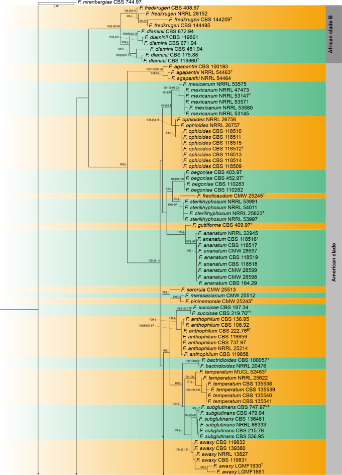
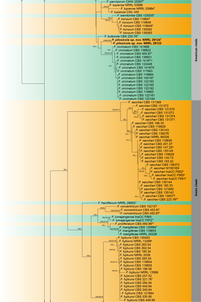
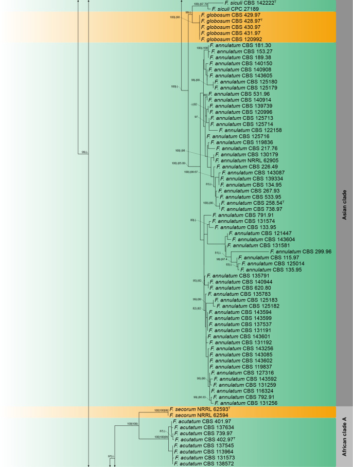
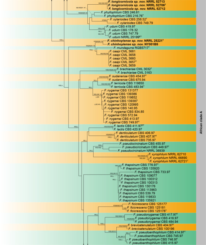
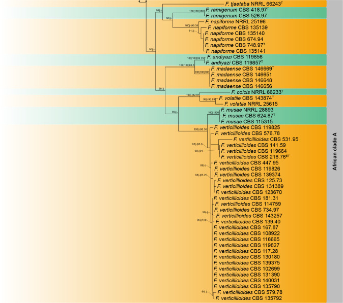
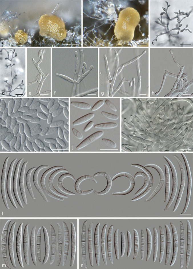
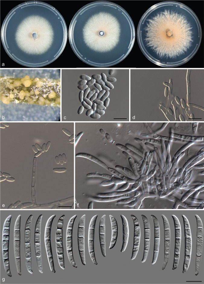
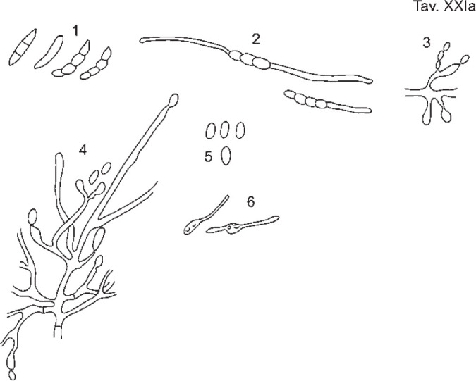
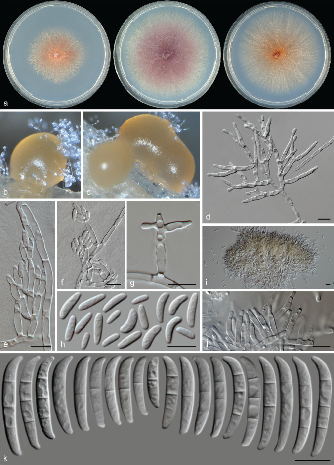
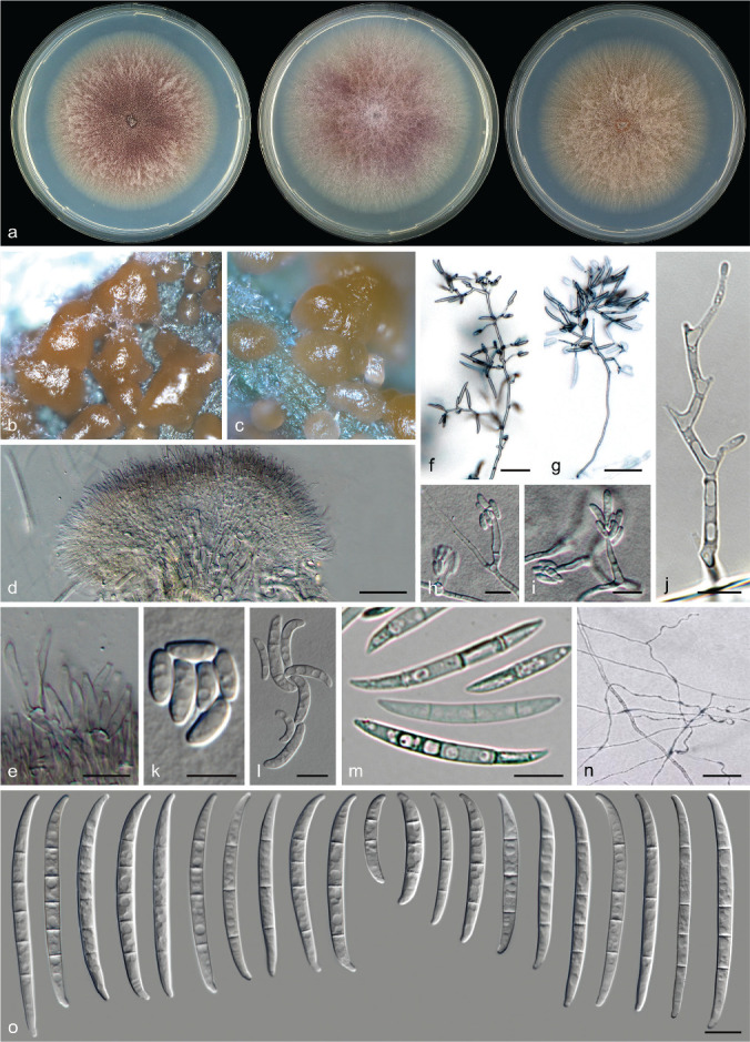
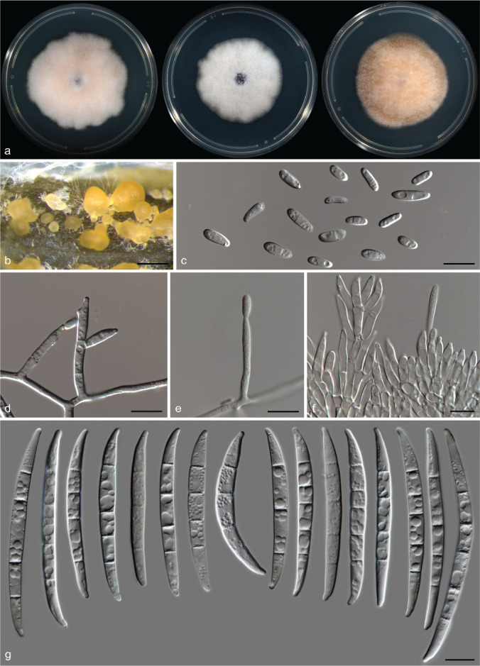
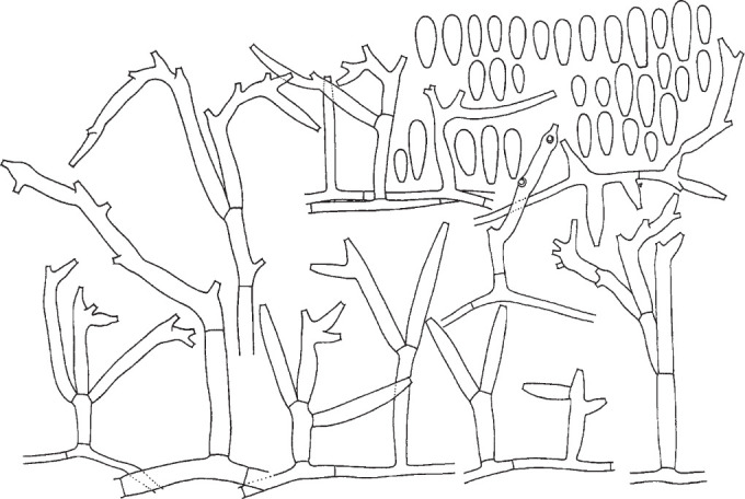
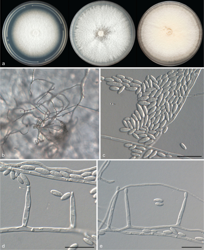
The Fusarium fujikuroi species complex (FFSC) includes more than 60 phylogenetic species (phylospecies) with both phytopathological and clinical importance. Because of their economical relevance, a stable taxonomy and nomenclature is crucial for species in the FFSC. To attain this goal, we examined type specimens and representative cultures of several species by employing morphology and phylogenetic analyses based on partial gene fragments of the translation elongation factor 1-alpha (tef1), beta-tubulin (tub2), calmodulin (cmdA), RNA polymerase largest subunit (rpb1) and RNA polymerase II second largest subunit (rpb2). Based on these results three new species were delimited in the FFSC. Two of these phylospecies clustered within the African clade, and one in the American clade. Epitypes were also designated for six previously described FFSC species including F. proliferatum and F. verticillioides, and a neotype designated for F. subglutinans. Furthermore, both F. acutatum and F. ophioides, which were previously invalidly published, are validated. Citation: Yilmaz N, Sandoval-Denis M, Lombard L, et al. 2021. Redefining species limits in the Fusarium fujikuroi species complex. Persoonia 46: 129-162. https://doi.org/10.3767/persoonia.2021.46.05.

Keywords: epitypification; fungal taxonomy; morphology; neotypification; new taxa; validation.


Figures

Fig. 1
Fig. 1
Combined phylogeny of the tef1, rpb2, rpb1, tub2 and cmdA gene regions of species from Fusarium fujikuroi species complex. Fusarium nirenbergiae (CBS 744.97) was selected as out-group. Strains belonging to new species are indicated in bold. Numbers at the branches indicate support values (bootstrap|gCF|sCF) above 80 %. T =.Ex-type, NT = neotype, ET = epitype. aEx-type of F. neoceras (CBS 147.25), bIsolates previously described as F. desaboruense (Maryani et al. 2019b).
Fig. 1
Fig. 1
Combined phylogeny of the tef1, rpb2, rpb1, tub2 and cmdA gene regions of species from Fusarium fujikuroi species complex. Fusarium nirenbergiae (CBS 744.97) was selected as out-group. Strains belonging to new species are indicated in bold. Numbers at the branches indicate support values (bootstrap|gCF|sCF) above 80 %. T =.Ex-type, NT = neotype, ET = epitype. aEx-type of F. neoceras (CBS 147.25), bIsolates previously described as F. desaboruense (Maryani et al. 2019b).
Fig. 2
Fig. 2
Fusarium annulatum (a, h, j–l. CBS 258.54T; b, c, g, i, m. CBS 531.96; d, f. CBS 139379; e. CBS 134.95; n. CBS 143601). a–b. Sporodochia formed on the surface of carnation leaves; c–h. aerial conidiophores and conidiogenous cells; i–j. aerial conidia; k. sporodochial conidiophores and conidiogenous cells; l–n. sporodochial conidia. — Scale bars: a–d = 50 μm; all others = 10 μm (scale bar in l also applies to m and n).
Fig. 3
Fig. 3
Fusarium chinhoyiense sp. nov. (NRRL 25221T). a. Colonies in PDA after 7 d at 25 °C light, dark and nuv (from left to right), respectively; b. sporodochia formed on the surface of carnation leaves; c. aerial conidia; d–e. aerial conidiophores and phialides; f. sporodochial conidiophores and phialides; g. sporodochial conidia. — Scale bars: c–g = 10 μm.
Fig. 4
Fig. 4
Lectotype of Fusarium lactis (Pirotta & Riboni 1879).
Fig. 5
Fig. 5
Fusarium longicornicola sp. nov. (NRRL 52706T). a. Colonies in PDA after 7 d at 25 °C light, dark and nuv (from left to right), respectively; b–c. sporodochia formed on the surface of carnation leaves; d–g. aerial conidiophores and phialides; h. aerial conidia; i–j. sporodochial conidiophores and phialides; k. sporodochial conidia. — Scale bars: d–k = 10 μm.
Fig. 6
Fig. 6
Fusarium ophioides (CBS 118512T). a. Colonies in PDA after 7 d at 25 °C light, dark and nuv (from left to right), respectively; b–c. sporodochia formed on the surface of carnation leaves; d–e. sporodochial conidiophores and phialides; f–j. aerial conidiophores and conidiogenous cells; k–l. microconidia; m. mesoconidia; n. serpentine hyphae (adapted from Jacobs 2010); o. sporodochial conidia. — Scale bars: d, f–g = 50 μm; k, n = 5 μm; all others = 10 μm.
Fig. 7
Fig. 7
Fusarium pilosicola sp. nov. (NRRL 29124T). a. Colonies in PDA after 7 d at 25 °C light, dark and nuv (from left to right), respectively; b. sporodochia formed on the surface of carnation leaves; c. aerial conidia; d–e. aerial conidiophores and phialides; f. sporodochial conidiophores and phialides; g. sporodochial conidia. — Scale bars: c–g = 10 μm.
Fig. 8
Fig. 8
Lectotype of Fusarium proliferatum (Matsushima 1971).
Fig. 9
Fig. 9
Fusarium proliferatum (CBS 480.96ET). a. Colonies in PDA after 7 d at 25 °C light, dark and nuv (from left to right), respectively; b. sporodochia formed on the surface of carnation leaves; c. aerial conidia; d–e. aerial conidiophores and phialides; f–g. sporodochial conidiophores and phialides; h. sporodochial conidia. — Scale bars: c–h = 10 μm.
Fig. 10
Fig. 10
Lectotype of Fusarium sacchari (Butler & Khan 1913).
Fig. 11
Fig. 11
Fusarium subglutinans (CBS 747.97ET). a. Colonies in PDA after 7 d at 25 °C light, dark and nuv (from left to right), respectively; b. sporodochia formed on the surface of carnation leaves; c. aerial conidia; d–e. aerial conidia, conidiophores and phialides; f. sporodochial conidiophores and phialides; g. sporodochial conidia. — Scale bars: c–g = 10 μm.
Fig. 12
Fig. 12
Lectotype of Fusarium verticillioides (Saccardo 1881).
Fig. 13
Fig. 13
Fusarium verticillioides (CBS 218.76ET). a. Colonies in PDA after 7 d at 25 °C light, dark and nuv (from left to right), respectively; b. aerial conidia produced in chains; c. aerial conidia; d–e. aerial conidia, conidiophores and phialides. — Scale bars: c–g = 10 μm.

Similar articles

Cited by

References

    1. Akaike H. 1974. A new look at the statistical model identification. IEEE Transations on Automatic Control 19: 716–723.
    1. Al-Hatmi AMS, Hagen F, Menken SBJ, et al. . 2016a. Global molecular epidemiology and genetic diversity of Fusarium, a significant emerging group of human opportunists from 1958 to 2015. Emerging Microbes & Infections 5: e124. - PMC - PubMed
    1. Al-Hatmi AMS, Mirabolfathy M, Hagen F, et al. . 2016b. DNA barcoding, MALDI-TOF, and AFLP data support Fusarium ficicrescens as a distinct species within the Fusarium fujikuroi species complex. Fungal Biology 120: 265–278. - PubMed
    1. Al-Hatmi AMS, Sandoval-Denis M, Nabet C, et al. . 2019. Fusarium volatile a new potential pathogen from a human respiratory sample. Fungal Systematics and Evolution 4: 171–181. - PMC - PubMed
    1. Aoki T, O’Donnell K, Geiser DM. 2014. Systematics of key phytopathogenic Fusarium species: current status and future challenges. Journal of General Plant Pathology 80: 189–201.

LinkOut - more resources