. 2022 Jul 19:13:939213.
doi: 10.3389/fimmu.2022.939213. eCollection 2022.

Low-dose mycophenolate mofetil improves survival in a murine model of Staphylococcus aureus sepsis by increasing bacterial clearance and phagocyte function

Affiliations

Low-dose mycophenolate mofetil improves survival in a murine model of Staphylococcus aureus sepsis by increasing bacterial clearance and phagocyte function

Fanny Alby-Laurent et al. Front Immunol. .

Abstract

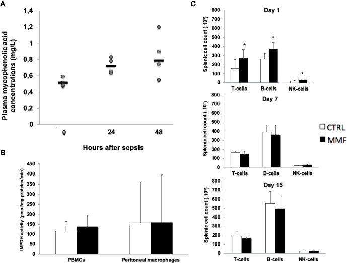
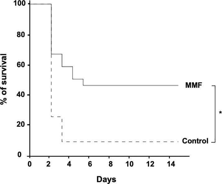
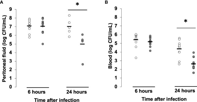
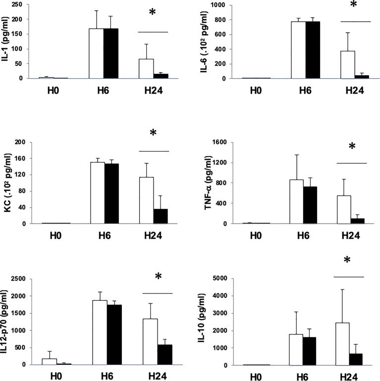
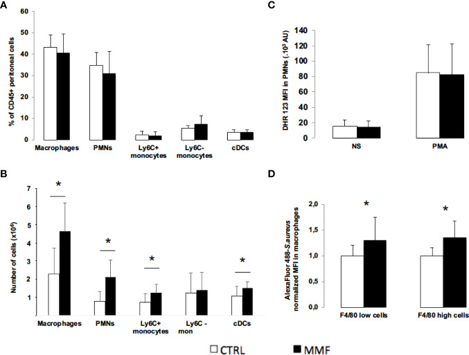
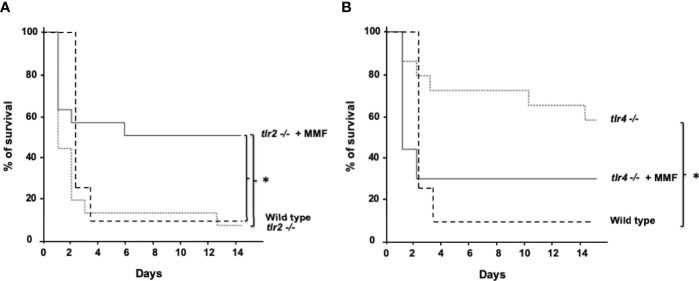
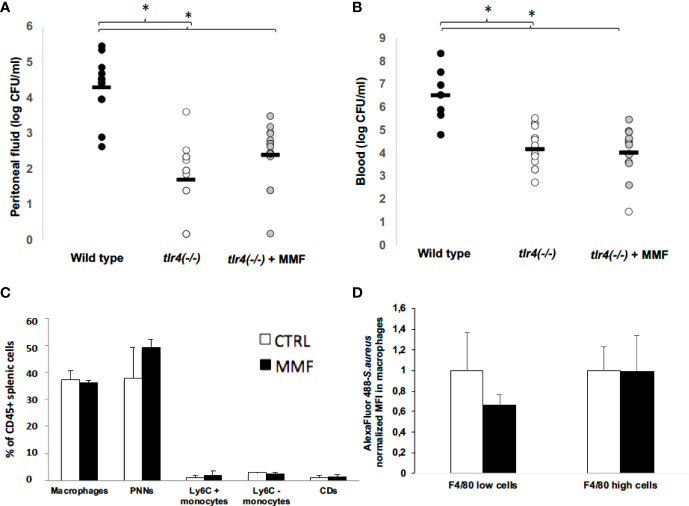
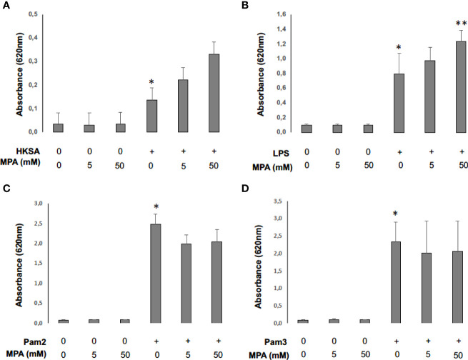
Regulators of TLRs signaling pathways play an important role in the control of the pro-inflammatory response that contributes to sepsis-induced tissue injury. Mycophenolate mofetil, an immunosuppressive drug inhibiting lymphocyte proliferation, has been reported to be a regulator of TLRs signaling pathways. Whether MMF used at infra-immunosuppressive doses has an impact on survival and on innate immune response in sepsis is unknown. C57BL/6J mice were infected intraperitoneally with 108 CFU Staphylococcus aureus, and treated or not with low-dose of MMF (20mg/kg/day during 4 days). Survival rate and bacterial clearance were compared. Cytokine levels, quantitative and qualitative cellular responses were assessed. S. aureus - infected mice treated with MMF exhibited improved survival compared to non-treated ones (48% vs 10%, p<0.001). With the dose used for all experiments, MMF did not show any effect on lymphocyte proliferation. MMF treatment also improved local and systemic bacterial clearance, improved phagocytosis activity of peritoneal macrophages resulting in decreased inflammatory cytokines secretion. MMF-treated mice showed enhanced activation of NF-κB seemed with a suspected TLR4-dependent mechanism. These results suggest that infra-immunosuppressive doses of MMF improve host defense during S. aureus sepsis and protects infected mice from fatal outcome by regulating innate immune responses. The signaling pathways involved could be TLR4-dependent. This work brings new perspectives in pathogenesis and therapeutic approaches of severe infections.

Keywords: NF-κB; innate immunity; macrophages; mycophenolate mofetil; phagocytosis; sepsis; staphylococcus aureus; toll-like receptor 4.


Conflict of interest statement

The authors declare that the research was conducted in the absence of any commercial or financial relationships that could be construed as a potential conflict of interest.

Figures

Figure 1
Figure 1
Low-dose MMF did not have any impact on splenic lymphocytes sub-population and IMDH activity during sepsis Mice received MMF i.p. 20 mg/kg once daily, one day before i.p. injection of 108 S. aureus and for 4 more days. Trough concentrations were measured at H0 (n=4), H12 (n=4) and H24 (n=5) (A). IMPDH activity was measured in PBMCs and in peritoneal macrophages 4 days after treatment with MMF (or vehicle), one hour after the last injection (B). Splenic lymphocyte sub-populations were counted by flow cytometry, at day 1, 7 and 15 of sepsis (C) *P<0,05.
Figure 2
Figure 2
MMF improves survival during i.p. S.aureus infection. Mice were treated with MMF, which started one day before i.p. injection of 108 CFU S. aureus and for 3 more days (dashed line, n=25) and were compared to control mice treated with an equivalent volume of NaCl 0.9% (Dotted line, n=25). Survival was monitored for 14 days after infection. *Log-rank test, P<0.01.
Figure 3
Figure 3
MMF improves bacterial clearance in mice infected with S. aureus. Mice were treated with MMF, which started one day before i.p. injection of 108 CFU S. aureus (n=10, grey dots) and were compared to control mice treated with an equivalent volume of NaCl 0.9% (control, n=10, white dots). 6 and 24 hours after infection, mice were euthanized and quantitative bacterial culture was assessed in peritoneal fluid (A) and blood (B). Differences between MMF and control conditions were calculated using Student’s t test. * P<0.05.
Figure 4
Figure 4
MMF treatment attenuates the release of pro-inflammatory and anti-inflammatory cytokines during infection with S. aureus. Mice were treated with MMF, which started one day before i.p. injection of 108 CFU S. aureus (black histograms) and were compared to control mice treated with an equivalent volume of NaCl 0.9% (white histograms). Mice were euthanized 6 (n=5 in each group) and 24 hours (n=10 in each group) after infection. Serum IL-1, IL-6, KC, TNF, IL12p70 and IL-10 were measured by Mesoscale Discovery technology. Data are represented as means ± SD. Differences were calculated using Student’s t test. * P<0.05.
Figure 5
Figure 5
Effect of MMF on innate immune cells distribution and functions during S. aureus sepsis Mice were treated with MMF (n=10, black histogram), or with the same volume of NaCl 0.9% (control mice, n=10, white histogram). 24 hours after infection, peritoneal and splenic cells were stained for CD45, CD11b, Ly6C and Ly6G (PMNs), CD11b and Ly6C (monocytes), CD11b and F4/80 (macrophages) and CD11c (conventional dendritic cells) (A, B). Mice were treated with MMF (black histogram, n=14) or the same volume of NaCl 0.9% (white histogram, n=14) and were infected with S. aureus one hour after the 2nd injection. Peritoneal cells were collected 16 hours after infection, incubated with DHR 123 and cells were then activated in vitro by PMA (0.2 μg). NS represents non-stimulated condition. Peritoneal cells were stained for the PMN markers and DHR 123 fluorescence was measured by flow cytometry (C). Peritoneal cells (6. 105 cells) were isolated from mice treated with MMF (black histogram) or NaCl 0.9% (white histogram) for 4 days, and were incubated 30 minutes with S. aureus (MOI 10) stained with Alexa Fluor 488. Cells were then stained with F4/80, and intracellular MFI was measured by flow cytometry in F4/80+ macrophages and F4/80 high macrophages. MFI for each condition was normalized on the average of MFI in control mice for each experiment. Data were representative of 6 experiments (D). Data are represented as means ± SD. Differences were calculated using Student’s t test. * P<0.05.
Figure 6
Figure 6
MMF improves survival during i.p. S. aureus infection in Tlr2(-/-) mice (A) but not in Tlr4(-/-) mice (B) Tlr2(-/-) and Tlr4(-/-) mice were treated with MMF, which started one day before i.p. injection of 108 CFU S. aureus and for 3 more days (dashed line, n=17 and n=15, respectively, representing 4 experiments) and were compared to Tlr2(-/-) and Tlr4(-/-) control mice (Dotted line, n=17 and n=15 respectively, representing 4 experiments) and with wild type control mice treated with an equivalent volume of NaCl 0.9% (n=25, representing the 8 experiments). Survival was monitored for 14 days after infection. *Log-rank test, P<0.01.
Figure 7
Figure 7
MMF has no impact on bacterial clearance, on splenic and peritoneal cells and on macrophages phagocytosis activity 24 hours after S. aureus infection in Tlr4(-/-) mice Tlr4(-/-) mice. Mice were treated with MMF, which started one day before i.p. injection of 108 CFU S. aureus (n=14, grey dots) and were compared to control mice treated with an equivalent volume of NaCl 0.9% (wild type, n= 12, black dots; Tlr4(-/-) control mice, n=14, white dots). At H24 after infection, mice were euthanized and quantitative bacterial culture was assessed in peritoneal fluid (A) and blood (B). Mice were treated i.p. with MMF (n=7, black histogram), or with the same volume of NaCl 0.9% (control mice, n=7, white histogram), which started one day before the i.p. injection of S. aureus. 24 hours after infection, splenic cells were stained for CD45, CD11b, Ly6C and Ly6G (PMNs), CD11b and Ly6C (monocytes), CD11b and F4/80 (macrophages) and CD11c (conventional dendritic cells) (C). Peritoneal cells (6. 106 cells) were isolated from Tlr4 (-/-) mice treated with MMF (black histogram) or NaCl 0.9% (white histogram) for 4 days, and were incubated 30 minutes with S. aureus (MOI 10) stained with Alexa Fluor 488. Cells were then stained for the macrophage marker F4/80, and intracellular mean fluorescence of intensity (MFI) was measured by flow cytometry in F4/80+ macrophages and F4/80 high macrophages. MFI for each condition was normalized on the average of MFI in control mice for each experiment. Data were representative of 2 experiments (D). Differences between MMF and control conditions were calculated using ANOVA with Tukey’s multiple comparisons tests and Student’s t test. * P<0.05.
Figure 8
Figure 8
Mycophenolic acid potentiates NF-κB activity following monocyte cell-stimulation by S. aureus. THP1-Blue NF-kB cells (2. 105 cells) were incubated 24 hours at 37°C with CO2 5% with different concentrations of MPA and stimulated with HKSA (1 x 10⁵) (A), LPS (0.05ng/μl) (B), Pam2 (0,05ng/μl) (C) or Pam3 (0,05ng/μl) (D). Optical density (OD) at 620 nm reflecting NF-κB activity was then read with EPOCH spectrometry in 96-well-plate. Differences between non-stimulated and stimulated conditions and between stimulated conditions with and without MPA were calculated using ANOVA with Tukey’s multiple comparisons tests. * represents P<0.05 between non-stimulated and stimulated conditions, ** P<0.05 represents P<0.05 between stimulated conditions with and without MPA.

Similar articles

Cited by

References

    1. Singer M, Deutschman CS, Seymour CW, Shankar-Hari M, Annane D, Bauer M, et al. . The third international consensus definitions for sepsis and septic shock(sepsis-3). JAMA (2016) 315(8):801–10. doi: 10.1001/jama.2016.0287 - DOI - PMC - PubMed
    1. Angus DC, van der Poll T. Severe sepsis and septic shock. N Engl J Med (2013) 369:840–51. doi: 10.1056/NEJMra1208623 - DOI - PubMed
    1. Medzhitov R, Schneider DS, Soares MP. Disease tolerance as a defense strategy. Science (2012) 335:936–41. doi: 10.1126/science.1214935 - DOI - PMC - PubMed
    1. Hattori Y, Hattori K, Suzuki T, Matsuda N. Recent advances in the pathophysiology and molecular basis of sepsis-associated organ dysfunction: novel therapeutic implications and challenges. Pharmacol Ther (2017) 177:56–66. doi: 10.1016/j.pharmthera.2017.02.040 - DOI - PubMed
    1. Torgersen C, Moser P, Luckner G, Mayr V, Jochberger S, Hasibeder WR, et al. . Macroscopic postmortem findings in 235 surgical intensive care patients with sepsis. Anesth Analg (2009) 108:1841–7. doi: 10.1213/ane.0b013e318195e11d - DOI - PubMed

Publication types