

Hemodynamic and Geometric Risk Factors for In-Stent Restenosis in Patients with Intracranial Atherosclerotic Stenosis
- PMID: 35936215
- PMCID: PMC9348934
- DOI: 10.1155/2022/6951302
Hemodynamic and Geometric Risk Factors for In-Stent Restenosis in Patients with Intracranial Atherosclerotic Stenosis
Retraction in
-
Retracted: Hemodynamic and Geometric Risk Factors for In-Stent Restenosis in Patients with Intracranial Atherosclerotic Stenosis.Oxid Med Cell Longev. 2024 Jan 9;2024:9863972. doi: 10.1155/2024/9863972. eCollection 2024. Oxid Med Cell Longev. 2024. PMID: 38234560 Free PMC article.
Abstract
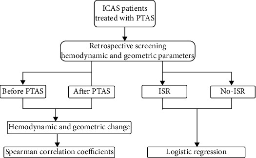
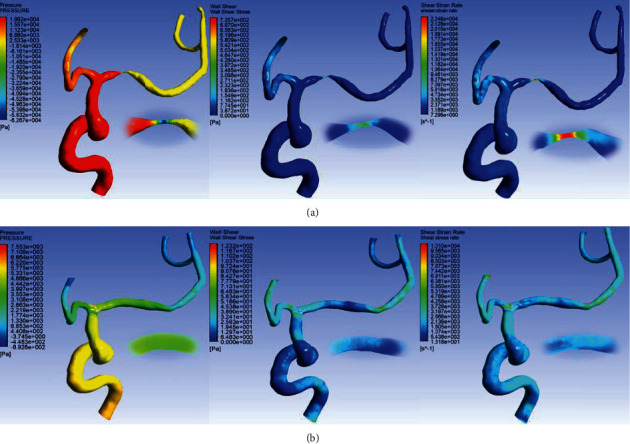
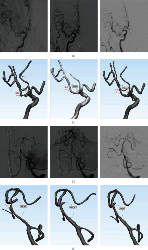
Methods: Severe ICAS patients managed with percutaneous transluminal angioplasty and stenting (PTAS) were included in the retrospective cohort study and were divided into two groups according to whether ISR occurred at follow-up (ISR group and no-ISR group). Computational fluid dynamics models were built based on digital subtraction angiography before and after PTAS to simulate blood flow and quantify hemodynamic parameters. The associations between vessel geometry, hemodynamics, and ISR in ICAS patients were investigated.
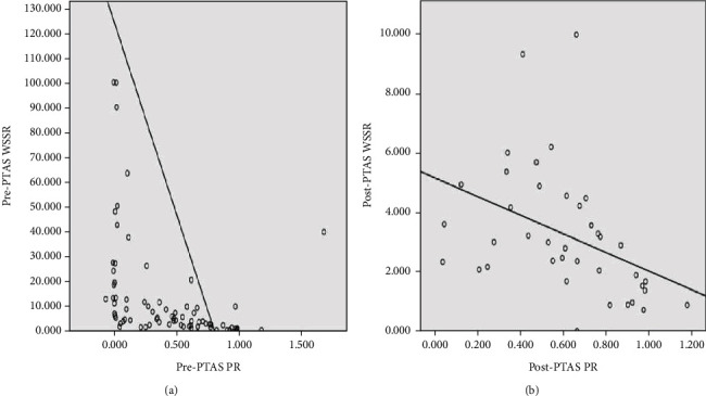
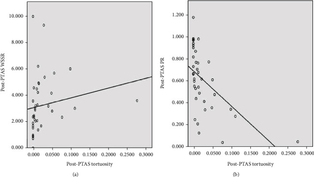
Results: Among 39 patients, ISR occurred in seven patients (17.95%) after a mean follow-up period of 6.69 ± 3.24 months. Stenting decreased vessel angulation (51.11° [40.07°-67.27°] vs. 15.97° [0.00°-36.16°], P = 0.000) and vessel tortuosity (0.09 [0.06-0.13] vs. 0.01 [0.00-0.03], P = 0.000). Meanwhile, the translational pressure ratio (PR) dramatically increased (0.07 [0.00-0.31] vs. 0.62 [0.41-0.82], P = 0.000) with the wall shear stress ratio decreased (13.93 [8.37-40.30] vs. 2.90 [1.69-4.48], P = 0.000). In the multivariate analysis, smaller Δ tortuosity (P = 0.038) was independently associated with the occurrence of ISR, and smaller post-PTAS translesional PR was also a predictive factor of marginal significance (P = 0.059).
Conclusion: PTAS decreased vessel angulation, vessel tortuosity, and translesional wall shear stress ratio while it increased translesional pressure ratio (PR) dramatically in ICAS patients. Smaller Δ tortuosity was found to be a risk factor for ISR, and smaller post-PTAS translesional PR was also a predictive factor of marginal significance, indicating that both geometric and hemodynamic parameters played important roles in the occurrence of ISR after PTAS.
Copyright © 2022 Xiaowen Song et al.
Conflict of interest statement
The authors declare that there is no conflict of interest regarding the publication of this article.
Figures
Similar articles
-
Peri-therapeutic multi-modal hemodynamic assessment and detection of predictors for symptomatic in-stent restenosis after percutaneous transluminal angioplasty and stenting.Front Neurol. 2023 Apr 18;14:1136847. doi: 10.3389/fneur.2023.1136847. eCollection 2023. Front Neurol. 2023. PMID: 37144006 Free PMC article.
-
Angiography-based hemodynamic features predict recurrent ischemic events after angioplasty and stenting of intracranial vertebrobasilar atherosclerotic stenosis.Eur Radiol. 2024 Apr;34(4):2352-2363. doi: 10.1007/s00330-023-10209-x. Epub 2023 Sep 19. Eur Radiol. 2024. PMID: 37723287 Free PMC article.
-
Baseline vessel wall magnetic resonance imaging characteristics associated with in-stent restenosis for intracranial atherosclerotic stenosis.J Neurointerv Surg. 2023 Mar;15(3):288-291. doi: 10.1136/neurintsurg-2021-018473. Epub 2022 Mar 1. J Neurointerv Surg. 2023. PMID: 35232754 Free PMC article.
-
Identifying risk factors for in-stent restenosis in symptomatic intracranial atherosclerotic stenosis: a systematic review and meta-analysis.Front Neurol. 2023 Jul 14;14:1170110. doi: 10.3389/fneur.2023.1170110. eCollection 2023. Front Neurol. 2023. PMID: 37521300 Free PMC article.
-
Clinical implications of haemodynamics in symptomatic intracranial atherosclerotic stenosis by computational fluid dynamics modelling: a systematic review.Stroke Vasc Neurol. 2025 Feb 25;10(1):16-24. doi: 10.1136/svn-2024-003202. Stroke Vasc Neurol. 2025. PMID: 38806205 Free PMC article.
Cited by
-
Has collateral blood flow any effect on restenosis rate? Our experience.Front Neurol. 2024 Feb 27;15:1360161. doi: 10.3389/fneur.2024.1360161. eCollection 2024. Front Neurol. 2024. PMID: 38476194 Free PMC article.
-
Cerebral hemodynamics in symptomatic anterior circulation intracranial stenosis measured by angiography-based quantitative flow ratio: association with CT perfusion.Eur Radiol. 2023 Aug;33(8):5687-5697. doi: 10.1007/s00330-023-09557-5. Epub 2023 Apr 6. Eur Radiol. 2023. PMID: 37022438
-
Retracted: Hemodynamic and Geometric Risk Factors for In-Stent Restenosis in Patients with Intracranial Atherosclerotic Stenosis.Oxid Med Cell Longev. 2024 Jan 9;2024:9863972. doi: 10.1155/2024/9863972. eCollection 2024. Oxid Med Cell Longev. 2024. PMID: 38234560 Free PMC article.
-
Evaluation of paclitaxel-coated balloon angioplasty for the treatment of symptomatic intracranial in-stent restenosis.Front Neurol. 2024 May 22;15:1360609. doi: 10.3389/fneur.2024.1360609. eCollection 2024. Front Neurol. 2024. PMID: 38841701 Free PMC article.
-
Complications and long-term in-stent restenosis of endovascular treatment of severe symptomatic intracranial atherosclerotic stenosis and relevant risk factors.Medicine (Baltimore). 2023 Sep 22;102(38):e34697. doi: 10.1097/MD.0000000000034697. Medicine (Baltimore). 2023. PMID: 37747021 Free PMC article.
References
-
- Derdeyn C. P., Chimowitz M. I., Lynn M. J., et al. Aggressive medical treatment with or without stenting in high-risk patients with intracranial artery stenosis (SAMMPRIS): the final results of a randomised trial. The Lancet . 2014;383(9914):333–341. doi: 10.1016/S0140-6736(13)62038-3. - DOI - PMC - PubMed
Publication types
MeSH terms
LinkOut - more resources
Full Text Sources
Medical
Research Materials
