

GABA tonic currents and glial cells are altered during epileptogenesis in a mouse model of Dravet syndrome
- PMID: 35936501
- PMCID: PMC9350930
- DOI: 10.3389/fncel.2022.919493
GABA tonic currents and glial cells are altered during epileptogenesis in a mouse model of Dravet syndrome
Erratum in
-
Corrigendum: GABA tonic currents and glial cells are altered during epileptogenesis in a mouse model of Dravet syndrome.Front Cell Neurosci. 2022 Oct 20;16:1066985. doi: 10.3389/fncel.2022.1066985. eCollection 2022. Front Cell Neurosci. 2022. PMID: 36339817 Free PMC article.
Abstract
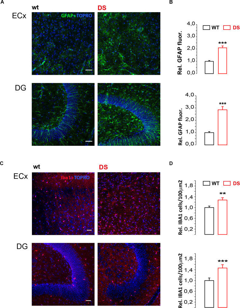
Dravet Syndrome (DS) is a rare autosomic encephalopathy with epilepsy linked to Nav1.1 channel mutations and defective GABAergic signaling. Effective therapies for this syndrome are lacking, urging a better comprehension of the mechanisms involved. In a recognized mouse model of DS, we studied GABA tonic current, a form of inhibition largely neglected in DS, in brain slices from developing mice before spontaneous seizures are reported. In neurons from the temporal cortex (TeCx) and CA1 region, GABA tonic current was reduced in DS mice compared to controls, while in the entorhinal cortex (ECx) it was not affected. In this region however allopregnanonole potentiation of GABA tonic current was reduced in DS mice, suggesting altered extrasynaptic GABAA subunits. Using THIP as a selective agonist, we found reduced δ subunit mediated tonic currents in ECx of DS mice. Unexpectedly in the dentate gyrus (DG), a region with high δ subunit expression, THIP-evoked currents in DS mice were larger than in controls. An immunofluorescence study confirmed that δ subunit expression was reduced in ECx and increased in DG of DS mice. Finally, considering the importance of neuroinflammation in epilepsy and neurodevelopmental disorders, we evaluated classical markers of glia activation. Our results show that DS mice have increased Iba1 reactivity and GFAP expression in both ECx and DG, compared to controls. Altogether we report that before spontaneous seizures, DS mice develop significant alterations of GABA tonic currents and glial cell activation. Understanding all the mechanisms involved in these alterations during disease maturation and progression may unveil new therapeutic targets.
Keywords: GFAP - glial fibrillary acidic protein; Iba-1; Nav1.1 channel; SCN1A encephalopathy; delta subunits; extrasynaptic GABA receptor A.
Copyright © 2022 Goisis, Chiavegato, Gomez-Gonzalo, Marcon, Requie, Scholze, Carmignoto and Losi.
Conflict of interest statement
The authors declare that the research was conducted in the absence of any commercial or financial relationships that could be construed as a potential conflict of interest.
Figures


Similar articles
-
Corrigendum: GABA tonic currents and glial cells are altered during epileptogenesis in a mouse model of Dravet syndrome.Front Cell Neurosci. 2022 Oct 20;16:1066985. doi: 10.3389/fncel.2022.1066985. eCollection 2022. Front Cell Neurosci. 2022. PMID: 36339817 Free PMC article.
-
Cyfip1 Haploinsufficiency Does Not Alter GABAA Receptor δ-Subunit Expression and Tonic Inhibition in Dentate Gyrus PV+ Interneurons and Granule Cells.eNeuro. 2019 Jun 28;6(3):ENEURO.0364-18.2019. doi: 10.1523/ENEURO.0364-18.2019. Print 2019 May/Jun. eNeuro. 2019. PMID: 31209152 Free PMC article.
-
Functional consequences of GABAA receptor alpha 4 subunit deletion on synaptic and extrasynaptic currents in mouse dentate granule cells.Alcohol Clin Exp Res. 2008 Jan;32(1):19-26. doi: 10.1111/j.1530-0277.2007.00564.x. Epub 2007 Dec 7. Alcohol Clin Exp Res. 2008. PMID: 18070250
-
Decreased surface expression of the δ subunit of the GABAA receptor contributes to reduced tonic inhibition in dentate granule cells in a mouse model of fragile X syndrome.Exp Neurol. 2017 Nov;297:168-178. doi: 10.1016/j.expneurol.2017.08.008. Epub 2017 Aug 16. Exp Neurol. 2017. PMID: 28822839 Free PMC article.
-
Tonic GABA inhibition in hippocampal dentate granule cells: its regulation and function in temporal lobe epilepsies.Acta Physiol (Oxf). 2013 Nov;209(3):199-211. doi: 10.1111/apha.12148. Epub 2013 Aug 8. Acta Physiol (Oxf). 2013. PMID: 23865761 Review.
Cited by
-
Astrocytes as a target for therapeutic strategies in epilepsy: current insights.Front Mol Neurosci. 2023 Jul 31;16:1183775. doi: 10.3389/fnmol.2023.1183775. eCollection 2023. Front Mol Neurosci. 2023. PMID: 37583518 Free PMC article. Review.
-
Emerging Insights into the Pathogenic Mechanisms of Dravet Syndrome.Neurochem Res. 2025 Jun 26;50(4):209. doi: 10.1007/s11064-025-04471-2. Neurochem Res. 2025. PMID: 40569358 Review.
-
Transcriptomic and electrophysiological alterations underlying phenotypic variability in SCN1A-associated febrile seizures.Sci Rep. 2025 Jul 10;15(1):24794. doi: 10.1038/s41598-025-09208-3. Sci Rep. 2025. PMID: 40634438 Free PMC article.
-
The Biallelic Inheritance of Two Novel SCN1A Variants Results in Developmental and Epileptic Encephalopathy Responsive to Levetiracetam.Biomedicines. 2024 Jul 31;12(8):1698. doi: 10.3390/biomedicines12081698. Biomedicines. 2024. PMID: 39200163 Free PMC article.
-
A Journey between Neurons and Astrocytes: A Tribute to Giorgio Carmignoto.Neurochem Res. 2025 Sep 4;50(5):282. doi: 10.1007/s11064-025-04527-3. Neurochem Res. 2025. PMID: 40906055
References
LinkOut - more resources
Full Text Sources
Molecular Biology Databases
Miscellaneous