. 2022 Jul 5:9:rbac044.
doi: 10.1093/rb/rbac044. eCollection 2022.

Proteomics reveals differential adsorption of angiogenic platelet lysate proteins on calcium phosphate bone substitute materials

Affiliations

Proteomics reveals differential adsorption of angiogenic platelet lysate proteins on calcium phosphate bone substitute materials

Richard da Costa Marques et al. Regen Biomater. .

Abstract

Protein adsorption on biomaterials for bone substitution, such as calcium phosphates (CaP), evokes biological responses and shapes the interactions of biomaterials with the surrounding biological environment. Proteins adsorb when CaP materials are combined with growth factor-rich hemoderivatives prior to implantation to achieve enhanced angiogenesis and stimulate new bone formation. However, the identification of the adsorbed proteins and their angiogenic effect on bone homeostasis remain incompletely investigated. In this study, we analyzed the adsorbed complex protein composition on CaP surfaces when using the hemoderivatives plasma, platelet lysate in plasma (PL), and washed platelet lysate proteins (wPL). We detected highly abundant, non-regenerative proteins and anti-angiogenic proteins adsorbed on CaP surfaces after incubation with PL and wPL by liquid chromatography and mass spectrometry (LC-MS) proteomics. Additionally, we measured a decreased amount of adsorbed pro-angiogenic growth factors. Tube formation assays with human umbilical endothelial cells demonstrated that the CaP surfaces only stimulate an angiogenic response when kept in the hemoderivative medium but not after washing with PBS. Our results highlight the necessity to correlate biomaterial surfaces with complex adsorbed protein compositions to tailor the biomaterial surface toward an enrichment of pro-angiogenic factors.

Keywords: angiogenesis; calcium phosphates; platelet lysate; protein adsorption; proteomics.


Figures

None
Graphical abstract
Figure 1.
Figure 1.
Principal workflow of protein source and adsorbed protein analysis. PLs were generated from human platelets in plasma and from washed platelets, respectively. Different bone substitutes were incubated with hemoderivative protein sources for protein adsorption. Incubated bone substitutes were washed three times with PBS, pH 7.4 to remove loosely bound and free proteins. Afterward, adsorbed proteins were recovered by a protein desorption step. Protein sources and adsorbed protein samples were analyzed by protein quantification, SDS-PAGE, silver staining and LC–MS. Tube formation assays were employed to study pro-angiogenic effects of hemoderivative protein sources and bone substitutes with adsorbed proteins. Protein crystal structures were taken from RCSB protein data bank. Images are not drawn to scale.
Figure 2.
Figure 2.
Human washed PL differs from plasma and PL in protein composition and growth factor concentration. (A) Characteristics of utilized hemoderivative protein sources. The main composition, source of material, preparation method, protein concentration (mg ml−1 ± SD, n = 4–8) and platelet (PLT) count are listed. Protein sources were analyzed by (B) SDS-PAGE with silver staining and (C) quantitative LC–MS proteomics. The bars indicate the percentage based on all identified proteins with protein annotation in nine different functional classes. (DF) Protein sources were evaluated for pro-angiogenic growth factors. The concentration of VEGF, FGF-2 and PDGF-AB was measured by ELISA (data are shown as mean ± SD, n (PDGF-AB, cP) = 2, n (others) = 4). The statistical significance was calculated by ANOVA with Tukey’s multiple comparison test (***P < 0.001, ****P < 0.0001); N/a, data not available.
Figure 3.
Figure 3.
Human wPL induces stronger angiogenic response in tube formation assays. (AC) The pro-angiogenic effect of hemoderivative protein sources was analyzed by employing tube formation assays with HUVECs. HUVECs were incubated with different concentrations of citrate plasma (cP), platelet lysate in plasma (PL) and lysate of washed platelets (wPL) on GeltrexTM LDEV-Free reduced growth factor basement membrane matrix for 18 h. For cP and PL different percentual dilutions (0.25–10%) in the cell culture medium were tested. For wPL different ratios of wPL in total volume (1:50–1:2) with cell culture medium were tested. Total tube length was evaluated with ImageJ and the plugin angiogenesis analyzer (−, negative control: medium without LVES; +, positive control: medium with LVES; data are shown as mean ± SD, n = 3–8). (D) Tube formation was analyzed with a comparable protein concentration of 1.1 mg/ml−1 for all three protein sources (data are shown as mean ± SD, n = 6–17). The statistical significance was calculated by ANOVA with Dunnett’s multiple comparison test (*P < 0.05, **P < 0.01, ***P < 0.001, ****P < 0.0001), performing the comparison with the negative control (A–C) or with wPL (D). (E) HUVECs on GeltrexTM after 18 h incubation with different hemoderivative protein sources at the same concentration, as seen in (D). The scale bars represent 200 µm.
Figure 4.
Figure 4.
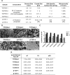
CaP Material characterization, SEM images and protein adsorption. (A) The physicochemical characterization of utilized CaP materials. The composition, size of the granules, crystal size, SSA, microporosity and the manufacturing processes are listed. Crystal size, SSA and microporosity for β-TCP materials were retrieved from previously published measurements [29]. (B) SEM images of granular CaP surfaces. The scale bars represent 10 µm. (C) Quantification of adsorbed proteins on CaPs, shown as a bar diagram. CaPs were incubated for 1 h in cP, PL and wPL, respectively. Protein adsorption of wPL on β-TCP 2 was not performed and analyzed due to limited quantities of wPL and strong similarities between β-TCP 1 and β-TCP 2 for cP and PL. Subsequently, three wash steps were performed and proteins were desorbed with 2% SDS. Proteins were quantified by Pierce 660 nm Assay (data are shown as mean ± SD, n = 2–7). (D) Quantification of adsorbed proteins on CaPs, shown as a table, as seen in (C) (data are shown as mean ± SD, n = 2–7).
Figure 5.
Figure 5.
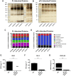
Cytoskeletal proteins from human washed PL are enriched on CaP surfaces while pro-angiogenic growth factors are depleted. CaP surfaces were incubated for 1 h in the hemoderivative protein source. After three subsequent washing steps, proteins were desorbed with 2% SDS. (A, B) SDS-PAGE and silver staining were performed for adsorbed proteins on different CaP surfaces incubated in PL and wPL, respectively. Protein adsorption of wPL on β-TCP 2 was not performed and analyzed due to limited quantities of wPL and strong similarities between β-TCP 1 and β-TCP 2 for cP and PL. (C, D) Adsorbed proteins on CaP surfaces from PL and wPL, respectively, were analyzed by quantitative LC–MS proteomics and identified proteins were classified into nine different protein functional classes. The bars indicate the percentage based on all identified proteins. (FG) Adsorbed proteins on the CaP surface of TCP/HA-4, incubated in wPL, were evaluated for angiogenesis-involved growth factors and compared with wPL. The concentration of VEGF, FGF-2 and PDGF-AB was measured by ELISA (b.d.l. = below detection limit; data are shown as mean ± SD, n = 4). The statistical significance was calculated by an unpaired t test (****P < 0.0001).
Figure 6.
Figure 6.
Non-regenerative and anti-angiogenic proteins are detected among the most abundant proteins on PL incubated CaP. (A, B) The heat maps show the 20 most abundant proteins for TCP/HA-4 compared with the other CaP surfaces. The values represent the percentage based on all identified proteins.
Figure 7.
Figure 7.
Washed CaP surfaces incubated in human washed PL show no pro-angiogenic effect. (A) The pro-angiogenic effect was analyzed by employing tube formation assays with HUVECs. HUVECs were incubated with untreated CaP surfaces and CaP surfaces, incubated in wPL and washed three times (all TCP/HA-4). HUVECs were then seeded on GeltrexTM LDEV-Free reduced growth factor basement membrane matrix for 18 h. Total tube length was evaluated with ImageJ and the plugin angiogenesis analyzer (−, negative control: medium without LVES; +, positive control: medium with LVES; data are shown as mean ± SD, n = 4–9). The statistical significance was calculated by ANOVA with Dunnett’s multiple comparison test (*P < 0.05), performing the comparison with the negative control. (B) HUVECs on GeltrexTM after 18 h incubation with untreated CaP surfaces and CaP surfaces (seen as dark spots in the images), incubated in wPL and washed three times, as seen in (A). The scale bars represent 200 µm.
Figure 8.
Figure 8.
CaP Surfaces kept in different hemoderivative protein source show a similar pro-angiogenic stimulation. (AC) The pro-angiogenic effect of CaP surfaces in hemoderivative protein sources was analyzed by employing tube formation assays with HUVECs. HUVECs were incubated with different concentrations of cP, PL and wPL, containing CaP on GeltrexTM LDEV-Free reduced growth factor basement membrane matrix for 18 h (all TCP/HA-4). for cP and PL different percentual dilutions (0.25–10%) in the cell culture medium were tested. For wPL different ratios of wPL in total volume (1:50–1:2) with cell culture medium were tested. Total tube length was evaluated with ImageJ and the plugin angiogenesis analyzer (−, negative control: medium without LVES; +, positive control: medium with LVES; data are shown as mean ± SD, n = 3–6). (D) Tube formation was analyzed with the same protein concentration of 1.1 mg/ml for all three protein sources, containing CaP without additional washing with PBS (all TCP/HA-4, data are shown as mean ± SD, n = 6–17). The statistical significance was calculated by ANOVA with Dunnett’s multiple comparison test (*P < 0.05, **P < 0.01, ***P < 0.001, ****P < 0.0001), performing the comparison with the negative control (A–C) or with wPL (D). (E) HUVECs on GeltrexTM after 18 h incubation with different hemoderivative protein sources at the same concentration, containing CaP (seen as dark spots in the images), as shown in (D). The scale bars represent 200 µm.

Similar articles

Cited by

References

    1. Horbett TA. Protein adsorption on biomaterials. In: Cooper SL, Peppas NA, Hoffman AS et al. (eds). Biomaterials, Interfacial Phenomena and applications. Washington, DC: American Chemical Society, 1982;233–44.
    1. Wang K, Zhou C, Hong Y, Zhang X.. A review of protein adsorption on bioceramics. Interface Focus 2012;2:259–77. - PMC - PubMed
    1. Horbett TA. The role of adsorbed proteins in tissue response to biomaterials. In: Ratner BD (ed). Biomaterials Science: An Introduction to Materials in Medicine. Amsterdam; Boston: Elsevier Academic Press, 2004;237–46.
    1. Bauer TW, Muschler GF.. Bone graft materials. An overview of the basic science. Clin Orthop Relat Res 2000;371:10–27. - PubMed
    1. Kurien T, Pearson RG, Scammell BE.. Bone graft substitutes currently available in orthopaedic practice: the evidence for their use. Bone Joint J 2013;95-B:583–97. - PubMed