

Label-Free Delineation of Human Uveal Melanoma Infiltration With Pump-Probe Microscopy
- PMID: 35936703
- PMCID: PMC9354715
- DOI: 10.3389/fonc.2022.891282
Label-Free Delineation of Human Uveal Melanoma Infiltration With Pump-Probe Microscopy
Abstract
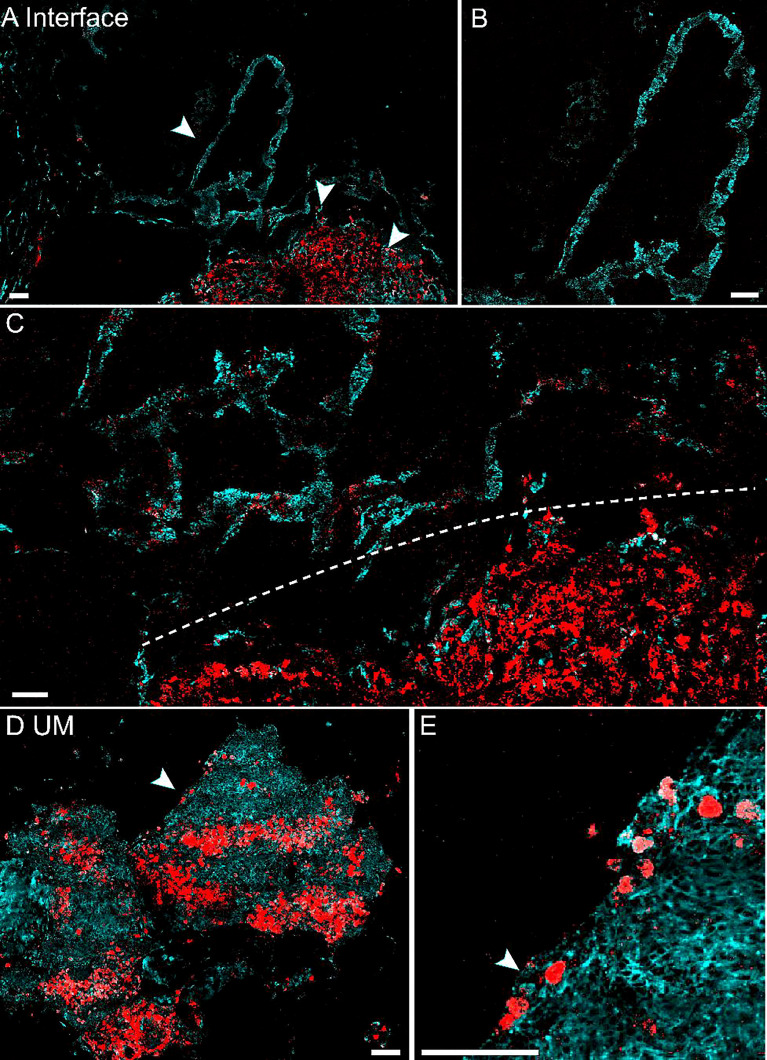
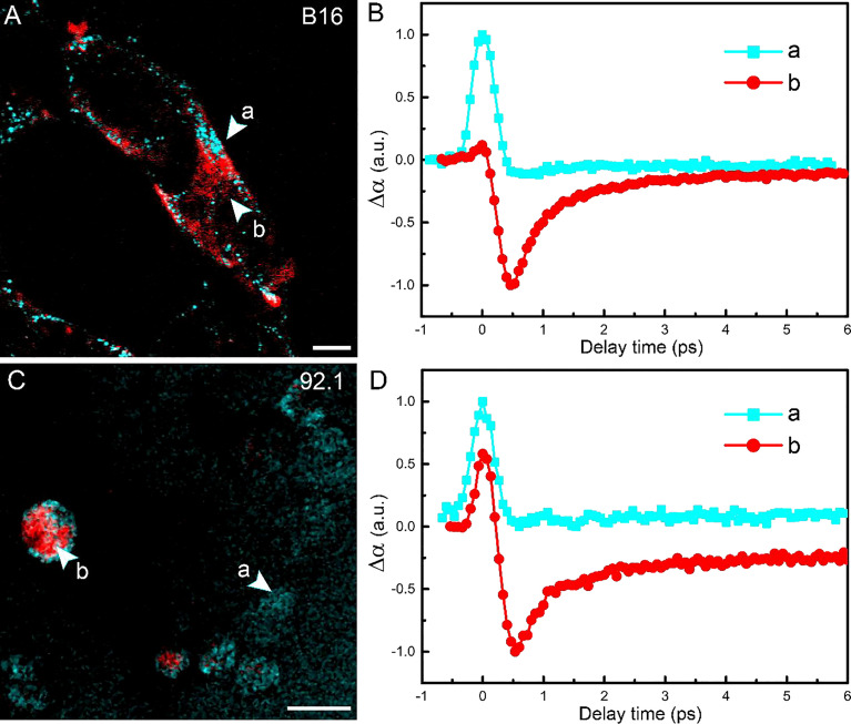
Uveal melanoma (UM) is the most frequent primary intraocular malignancy in adults, characterized by melanin depositions in melanocytes located in the uveal tract in the eyes. Differentiation of melanin species (eumelanin and pheomelanin) is crucial in the diagnosis and management of UM, yet it remains inaccessible for conventional histology. Here, we report that femtosecond time-resolved pump-probe microscopy could provide label-free and chemical-specific detection of melanin species in human UM based on their distinct transient relaxation dynamics at the subpicosecond timescale. The method is capable of delineating the interface between melanoma and paracancerous regions on various tissue conditions, including frozen sections, paraffin sections, and fresh tissues. Moreover, transcriptome sequencing was conducted to confirm the active eumelanin synthesis in UM. Our results may hold potential for sensitive detection of tumor boundaries and biomedical research on melanin metabolism in UM.
Keywords: label-free imaging; melanin; multiphoton microscopy; pump-probe microscopy; uveal melanoma.
Copyright © 2022 Zhang, Yao, Chen, Wang, Bao, Wang, Zhao and Ji.
Conflict of interest statement
The authors declare that the research was conducted in the absence of any commercial or financial relationships that could be construed as a potential conflict of interest. The handling editor ML and the reviewer LQ declared a shared parent affiliation with the authors BZ, YC, and MJ at the time of review.
Figures


Similar articles
-
Comparison of eumelanin and pheomelanin content between cultured uveal melanoma cells and normal uveal melanocytes.Melanoma Res. 2009 Apr;19(2):75-9. doi: 10.1097/CMR.0b013e328329ae49. Melanoma Res. 2009. PMID: 19262410
-
Characterization of melanin in human iridal and choroidal melanocytes from eyes with various colored irides.Pigment Cell Melanoma Res. 2008 Feb;21(1):97-105. doi: 10.1111/j.1755-148X.2007.00415.x. Pigment Cell Melanoma Res. 2008. PMID: 18353148
-
Imaging microscopic pigment chemistry in conjunctival melanocytic lesions using pump-probe laser microscopy.Invest Ophthalmol Vis Sci. 2013 Oct 21;54(10):6867-76. doi: 10.1167/iovs.13-12432. Invest Ophthalmol Vis Sci. 2013. PMID: 24065811 Free PMC article.
-
Uveal melanoma: A pathologist's perspective and review of translational developments.Adv Anat Pathol. 2014 Mar;21(2):138-43. doi: 10.1097/PAP.0000000000000010. Adv Anat Pathol. 2014. PMID: 24508696 Review.
-
Proteomics in uveal melanoma research: opportunities and challenges in biomarker discovery.Expert Rev Proteomics. 2007 Apr;4(2):273-86. doi: 10.1586/14789450.4.2.273. Expert Rev Proteomics. 2007. PMID: 17425462 Review.
References
LinkOut - more resources
Full Text Sources