

doi: 10.1016/j.eats.2022.02.026.
eCollection 2022 Jul.
Meniscal Ramp Repair: A 2-Portal Posteromedial Approach
Affiliations
- PMID: 35936835
- PMCID: PMC9353068
- DOI: 10.1016/j.eats.2022.02.026
Item in Clipboard
Meniscal Ramp Repair: A 2-Portal Posteromedial Approach
Arthrosc Tech.
.
Abstract
The management of medial meniscus ramp lesions can be challenging. The current gold standard technique to repair these lesions is the transnotch view combined with a single instrumental posteromedial portal. However, it does not provide direct visualization of the ramp and does not allow for an anatomic repair. In this Technical Note, a new technique is described with 2 posteromedial portals: a posteromedial viewing portal and working portal. This 2-portal approach aims to improve visualization of the lesion and its repair, as well as allow for a technically easier repair.
© 2022 The Authors.
Figures

Typical MRI sign of a ramp lesion in an ACL-deficient knee. In this sagittal slice MRI of the right knee, a hyper T2-weighted signal can be observed at the meniscocapsular junction of the posterior part of the medial meniscus posterior horn. Abbreviations: ACL, anterior cruciate ligament; MCJ, meniscocapsular junction; MM, medial meniscus; MRI, magnetic resonance imaging.
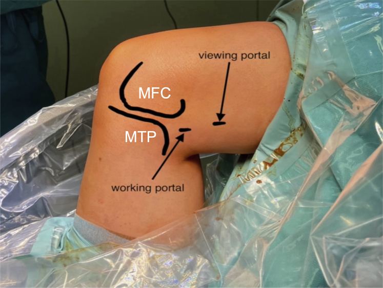
Landmarks and position of the posteromedial portals. The patient is lying supine on the operating table with the operated limb hanging toward the floor in a leg holder at 90° knee flexion. In this right knee, the viewing portal is posterior to the posterior femoral cortex and proximal to the posterior border of the medial femoral condyle. The working portal is located 3 to 4 cm more distal and slightly posterior to the viewing portal, at the height of the joint line. Abbreviations: MFC, medial femoral condyle; MTP, medial tibial plateau.

Viewing portal approach via the transnotch view. This is a right knee at 90° knee flexion (A). The entry of the viewing portal is identified through a transnotch articular view with the help of a needle and transillumination to protect from saphenous vessel injury (B). A no. 11 blade scalpel is used for the skin incision (C). Abbreviations: MFC, medial femoral condyle; MM, medial meniscus; PC, posterior capsule.
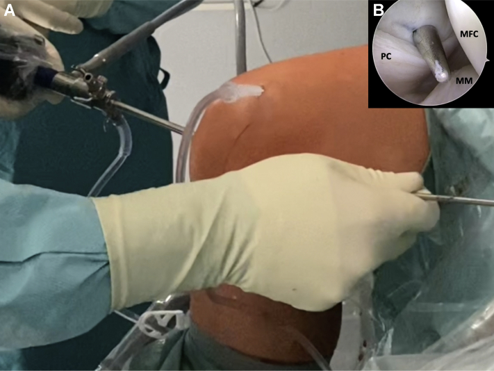
The use of a switching stick to help the introduction of the trocar and the camera into the posteromedial viewing portal. This a right knee at 90° flexion (A). After skin incision and dissection with scissors at the posteromedial viewing portal, a switching stick is used to help introduce the trocar and the camera into the posteromedial viewing portal (B). Abbreviations: MM, medial meniscus; PC, posterior capsule.
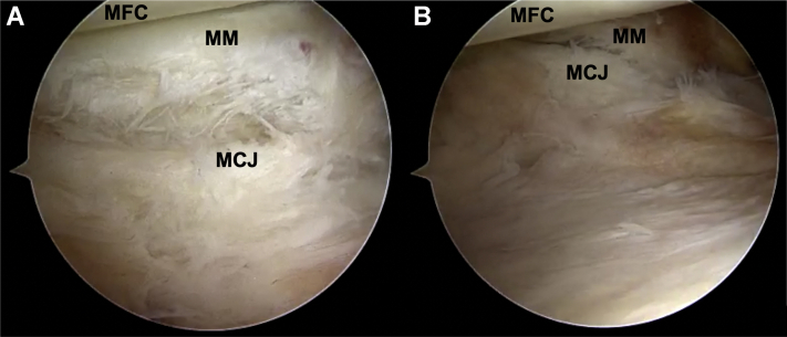
Direct visualization of the posteromedial corner and a ramp lesion through the posteromedial viewing portal (right knee). The camera is in the posteromedial viewing portal. The flexion-extension movement of the knee helps to reveal ramp lesions. (A) Posteromedial view of a ramp lesion at 90° flexion. (B) Posteromedial view of a ramp lesion at 20° extension. Abbreviations: MCJ, meniscocapsular junction; MFC, medial femoral condyle; MM, medial meniscus.

Instrumental portal approach, right knee at 90° flexion. The camera is in the posteromedial viewing portal. The entry of the working portal is identified with a needle and the use of trans illumination to protect from saphenous vessel injury (A). A no. 11 blade scalpel is used for skin incision (B). The ramp lesion is probed (C) and then debrided with a shaver (D). Abbreviations: MCJ, meniscocapsular junction; MFC, medial femoral condyle; MM, medial meniscus.

Suture of a ramp lesion with a 90° curved hook, right knee at 90° flexion. The arthroscopic view is posteromedial with the camera in the posteromedial viewing portal, and the instruments are introduced through the posteromedial instrumental portal. The joint capsule is grasped (A). The instrument is passed through the posterior meniscal border, and the PDS 0 suture is advanced in the posterior joint space (B). After suture retrieval, a sliding knot is made using a knot pusher (C). After section of the suture, the knot can be visualized (D). Abbreviations: MCJ, meniscocapsular junction; MFC, medial femoral condyle; MM, medial meniscus.

Posteromedial view before and after repair of a ramp lesion, right knee at 90° flexion. The camera is in the posteromedial viewing portal. Before repair, the ramp lesion is observed at 90° (A) and 20° (B) knee flexion. A cleft between the posterior wall of the medial meniscus and the ramp tissue can be identified in both positions. After repair, posteromedial view at 90° (A) and 20° (B) knee flexion. The blue star indicates adequate tensioning of the posterior capsule by the repair (B and D). Abbreviations: MCJ, meniscocapsular junction; MFC, medial femoral condyle; MM, medial meniscus.
References
-
- Greif D.N., Baraga M.G., Rizzo M.G., et al. MRI appearance of the different meniscal ramp lesion types, with clinical and arthroscopic correlation. Skeletal Radiol. 2020;49:677–689. - PubMed
-
- Kunze K.N., Wright-Chisem J., Polce E.M., DePhillipo N.N., LaPrade R.F., Chahla J. Risk factors for ramp lesions of the medial meniscus: A systematic review and meta-analysis. Am J Sports Med. 2021;49:3749–3757. - PubMed
-
- Sonnery-Cottet B., Conteduca J., Thaunat M., Gunepin F.X., Seil R. Hidden lesions of the posterior horn of the medial meniscus: A systematic arthroscopic exploration of the concealed portion of the knee. Am J Sports Med. 2014;42:921–926. - PubMed
-
- Mouton C., Magosch A., Pape D., Hoffmann A., Nührenbörger C., Seil R. Ramp lesions of the medial meniscus are associated with a higher grade of dynamic rotatory laxity in ACL-injured patients in comparison to patients with an isolated injury. Knee Surg Sports Traumatol Arthrosc. 2020;28:1023–1028. - PubMed
-
- Peltier A., Lording T., Maubisson L., Ballis R., Neyret P., Lustig S. The role of the meniscotibial ligament in posteromedial rotational knee stability. Knee Surg Sports Traumatol Arthrosc. 2015;23:2967–2973. - PubMed
LinkOut - more resources
Full Text Sources
