

Bubble Sign: An Arthroscopic Technical Trick to Differentiate Between Partial- and Full-Thickness Rotator Cuff Tears
- PMID: 35936860
- PMCID: PMC9353585
- DOI: 10.1016/j.eats.2022.03.022
Bubble Sign: An Arthroscopic Technical Trick to Differentiate Between Partial- and Full-Thickness Rotator Cuff Tears
Abstract
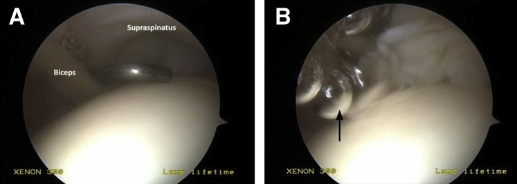
Distinguishing between partial-thickness and small focal full-thickness tears of rotator cuff may be important for determining the appropriate surgical treatment options and repair constructs in the care of patients with rotator cuff pathology. This article presents a simple intraoperative technical trick to aid in identification of small full-thickness tears of the superior rotator cuff. The relatively higher-pressured subacromial space and the low-pressured glenohumeral joint are separated by the supraspinatus tendon. When this barrier is compromised due to a full-thickness tear, free fluid flows from high to low pressure down the native pressure gradient. This is seen as the movement of air bubbles into the glenohumeral joint from the subacromial space and can be used to identify the presence of a full-thickness rotator cuff tear on diagnostic arthroscopy.
Figures

Comment in
-
Regarding "Bubble Sign: An Arthroscopic Technical Trick to Differentiate Between Partial- and Full-Thickness Rotator Cuff Tears".Arthroscopy. 2023 Mar;39(3):575. doi: 10.1016/j.arthro.2022.11.015. Arthroscopy. 2023. PMID: 36740281 No abstract available.
Similar articles
-
The supraspinatus distension sign: an indicator of supraspinatus integrity.Arthroscopy. 2009 Jun;25(6):617-9. doi: 10.1016/j.arthro.2008.11.009. Epub 2009 Jan 24. Arthroscopy. 2009. PMID: 19501291
-
Isolated Bioinductive Arthroscopic Repair of Partial-Thickness Rotator Cuff Tears Using a Resorbable Collagen Implant.JBJS Essent Surg Tech. 2022 Jan 25;12(1):e21.00008. doi: 10.2106/JBJS.ST.21.00008. eCollection 2022 Jan-Mar. JBJS Essent Surg Tech. 2022. PMID: 35685236 Free PMC article.
-
Biomechanical analysis of articular-sided partial-thickness rotator cuff tear and repair.Am J Sports Med. 2015 Feb;43(2):439-46. doi: 10.1177/0363546514560156. Epub 2014 Dec 15. Am J Sports Med. 2015. PMID: 25512665
-
Rotator cuff tears in overhead athletes.Clin Sports Med. 2012 Oct;31(4):675-92. doi: 10.1016/j.csm.2012.07.005. Clin Sports Med. 2012. PMID: 23040553 Review.
-
Surgery for rotator cuff tears.Cochrane Database Syst Rev. 2019 Dec 9;12(12):CD013502. doi: 10.1002/14651858.CD013502. Cochrane Database Syst Rev. 2019. PMID: 31813166 Free PMC article.
Cited by
-
Functional and MRI Outcomes After In Situ Repair Versus Tear Completion Before Repair of Bursal-Side Partial-Thickness Rotator Cuff Tears.Orthop Surg. 2023 Aug;15(8):2082-2090. doi: 10.1111/os.13693. Epub 2023 Mar 2. Orthop Surg. 2023. PMID: 36864555 Free PMC article.
References
-
- Schaeffeler C., Mueller D., Kirchhoff C., Wolf P., Rummeny E.J., Woertler K. Tears at the rotator cuff footprint: Prevalence and imaging characteristics in 305 MR arthrograms of the shoulder. Eur Radiol. 2011;21:1477–1484. - PubMed
-
- Guild T., Kuhn G., Rivers M., Cheski R., Trenhaile S., Izquierdo R. The role of arthroscopy in painful shoulder arthroplasty: Is revision always necessary? Arthroscopy. 2020;36:1508–1514. - PubMed
LinkOut - more resources
Full Text Sources
