

Point-of-care contrast enhanced lung ultrasound and COVID-19
- PMID: 35936970
- PMCID: PMC9354177
- DOI: 10.1177/1742271X211047945
Point-of-care contrast enhanced lung ultrasound and COVID-19
Abstract
Objectives: Bedside lung ultrasound has been indispensable during the coronavirus disease 2019 (COVID-19) pandemic, allowing us to rapidly assess critically unwell patients. We demonstrate the unique application of contrast-enhanced ultrasound with the aim of further understanding this disease.
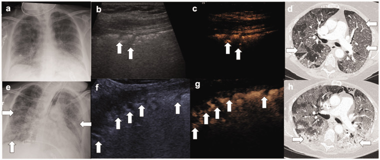
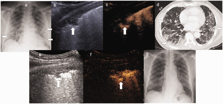
Methods: Patient demographics were recorded alongside recent cross-sectional imaging and inflammatory markers. Ultrasound was conducted by experienced operators in a portable setting. Conventional six-point lung ultrasound method was used to evaluate B-lines, small (subpleural) consolidation and the pleura. Areas of small consolidation were targeted after intravenous administration of ultrasound contrast.
Results: The areas of small consolidations, a potential sign of pneumonia on B-mode lung ultrasound, usually enhance on contrast-enhanced ultrasound. Our study revealed these areas to be avascular, indicating an underlying thrombotic/infarction process. Findings were present in 100% of the patients we examined. We have also shown that the degree of infarction correlates with CT severity (r = 0.4) and inflammatory markers, and that these areas improve as patients recover.
Conclusions: We confirmed the theory of immune thrombus by identifying the presence of microthrombi in the lungs of 100% of our patients, despite 79% having had a recent negative CT pulmonary angiogram study. contrast-enhanced ultrasound can be utilised to add confidence to an uncertain COVID-19 diagnosis and for prognosticating and monitoring progress in confirmed COVID-19 patients. Contrast-enhanced ultrasound is clearly very different to CT, the gold standard, and while there are specific pathologies that can only be detected on CT, contrast-enhanced ultrasound has many advantages, most notability the ability to pick up microthrombi at the periphery of the lungs.
Keywords: Coronavirus, consolidation, microthrombi, computed tomography, bedside ultrasound, critical care, microbubble.
© The Author(s) 2021.
Figures
Similar articles
-
Contrast enhanced ultrasound (CEUS) to assess pleural pulmonal changes in severe COVID-19 infection: First results.Clin Hemorheol Microcirc. 2020;75(1):19-26. doi: 10.3233/CH-209005. Clin Hemorheol Microcirc. 2020. PMID: 32538830 Free PMC article.
-
Lung Ultrasound in COVID-19 Pneumonia: Correlations with Chest CT on Hospital admission.Respiration. 2020;99(7):617-624. doi: 10.1159/000509223. Epub 2020 Jun 22. Respiration. 2020. PMID: 32570265 Free PMC article.
-
Zombie Cruise Ship Virtual Escape Room for POCUS Pulmonary: Scan Your Way Out.J Educ Teach Emerg Med. 2022 Jul 15;7(3):SG1-SG23. doi: 10.21980/J8RM0M. eCollection 2022 Jul. J Educ Teach Emerg Med. 2022. PMID: 37465772 Free PMC article.
-
Lung Ultrasound May Support Diagnosis and Monitoring of COVID-19 Pneumonia.Ultrasound Med Biol. 2020 Nov;46(11):2908-2917. doi: 10.1016/j.ultrasmedbio.2020.07.018. Epub 2020 Jul 20. Ultrasound Med Biol. 2020. PMID: 32807570 Free PMC article. Review.
-
Thoracic imaging tests for the diagnosis of COVID-19.Cochrane Database Syst Rev. 2020 Sep 30;9:CD013639. doi: 10.1002/14651858.CD013639.pub2. Cochrane Database Syst Rev. 2020. Update in: Cochrane Database Syst Rev. 2020 Nov 26;11:CD013639. doi: 10.1002/14651858.CD013639.pub3. PMID: 32997361 Updated.
Cited by
-
Critical Advances for Democratizing Ultrasound Diagnostics in Human and Veterinary Medicine.Annu Rev Biomed Eng. 2024 Jul;26(1):49-65. doi: 10.1146/annurev-bioeng-110222-095229. Epub 2024 Jun 20. Annu Rev Biomed Eng. 2024. PMID: 38166185 Free PMC article. Review.
-
Accuracy of point-of-care ultrasound examination of the lung in primary care performed by general practitioners: a cross-sectional study.BMC Prim Care. 2025 Apr 8;26(1):99. doi: 10.1186/s12875-025-02802-4. BMC Prim Care. 2025. PMID: 40200132 Free PMC article.
References
LinkOut - more resources
Full Text Sources