. 2022 Dec:12:100202.
doi: 10.1016/j.jvacx.2022.100202. Epub 2022 Aug 3.

Secreted heat shock protein gp96-Ig and OX40L-Fc combination vaccine enhances SARS-CoV-2 Spike (S) protein-specific B and T cell immune responses

Affiliations

Secreted heat shock protein gp96-Ig and OX40L-Fc combination vaccine enhances SARS-CoV-2 Spike (S) protein-specific B and T cell immune responses

Laura Padula et al. Vaccine X. 2022 Dec.

Abstract

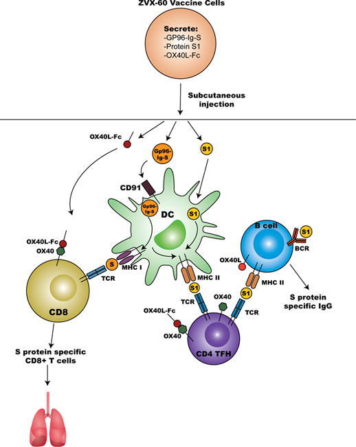
Encouraging protection results from current mRNA-based SARS-CoV-2 vaccine platforms are primarily due to the induction of SARS- CoV-2- specific B cell antibody and CD4 + T cell. Even though, current mRNA vaccine platforms are adept in inducing SARS-CoV2-specific CD8 + T cell, much less is known about CD8 T cells contribution to the overall vaccine protection. Our allogeneic cellular vaccine, based on a secreted form of the heat-shock protein gp96-Ig, achieves high frequencies of polyclonal CD8 + T cell responses to tumor and infectious antigens through antigen cross-priming in vivo. We and others have shown that gp96-Ig, in addition to antigen-specific CD8 + T cell anti-tumor and anti-pathogen immunity, primes antibody responses as well. Here, we generated a cell-based vaccine that expresses SARS-Cov-2 Spike (S) protein and simultaneously secretes gp96-Ig and OX40L-Fc fusion proteins. We show that co-secretion of gp96-Ig-S peptide complexes and the OX40L-Fc costimulatory fusion protein in allogeneic cell lines results in enhanced activation of S protein-specific IgG antibody responses. These findings were further strengthened by the observation that this vaccine platform induces T follicular helper cells (TFH) and protein-S -specific CD8 + T cells. Thus, a cell-based gp96-Ig vaccine/OX40-L fusion protein regimen provides encouraging translational data that this vaccine platform induces pathogen-specific CD8+, CD4 + T and B cell responses, and may cohesively work as a booster for FDA-approved vaccines. Our vaccine platform can be rapidly engineered and customized based on other current and future pathogen sequences.

Keywords: Antibody; B cells; CD8 T cells; Gp96; Heat shock protein; OX40L; SARS-CoV-2 protein S; TFH cells; Vaccine.


Conflict of interest statement

NS is inventor on the patent application No 62/983,783 entitled “Immune-mediated coronavirus treatments”; NS is a member of Heat Biologics COVID-19 Advisory Board. MMS is the Vice President of Research; ED is the Executive Director of Research.; PJ is the Director of Business Development, all are employed by Heat Biologics, Inc. RJ is the CEO of Pelican Therapeutics, a subsidiary of Heat Biologics, Inc. MMS, ED, PJ, RJ, and KP hold stock options in Heat Biologics, Inc. The remaining authors declare that the research was conducted in the absence of any commercial or financial relationships that could be construed as a potential conflict of interest.

Figures

None
Graphical abstract
Fig. 1
Fig. 1
Characteristics of the cell line expressing gp96-Ig, SARS-CoV-2 Spike (S) protein and OX40L-Fc. Cell line (AD100) was transfected with plasmids encoding a) gp96-Ig and full length protein S and b) gp96-Ig, full length protein S and OX40L-Fc. c) Secreted gp96-Ig was measured in the cell supernatant by ELISA. One million cells were plated in 1 ml for 24 h. Purified gp96-Ig was used as standard d) Secreted OX40L-Fc was measured in the cell supernatant by ELISA. One million cells were plated in 1 ml for 24 h. Purified OX40L-Fc was used as standard e) SARS-CoV2 protein S expression was analyzed by immunofluorescence f) SARS-CoV2 protein S expression in supernatant was measured by ELISA. One million cells were plated in 1 ml for 48 h and purified SARS-CoV2 protein S was used as standard.
Fig. 2
Fig. 2
Gp96-Ig and OX40L-Fc increase S protein specific IgG responses in vivo. a) C57Bl6 mice were vaccinated at day 0 and 14 with different concentrations of cell-based gp96-Ig vaccine that expressed SARS-CoV-2 glycoprotein S and OX40L-Fc or with AD100 or PBS (controls). b) Mice were vaccinated at day 0 and 14 with 1 μg/ml of ZVX-55 and ZVX-60 or with AD100 and PBS (controls). Serum was collected 5 days after last vaccination, and S protein specific IgG response was analyzed by ELISA. c) Mice were vaccinated at day 0 and 14 with 1 μg/ml ZVX-55 or ZVX-60 and S protein specific IgG response in serum was analyzed at day 5, 14 and 19. Data represent 3 independent biological replicates per group and mean ± standard error. To compare control (ZVX55) with experimental (ZVX60) group (alpha level of 0.05) unpaired t-test (two-tailed) was applied, *p < 0.05, **p < 0.01, and ***p < 0.001.
Fig. 3
Fig. 3
Gp96-Ig and OX40L-Fc induce B cells responses. C57Bl6 mice were vaccinated at day 0 and 14 with a cell-based gp96-Ig vaccine that expressed SARS-CoV-2 glycoprotein S (ZVX-55, 1ug gp96-Ig) and OX40L-Fc (ZVX-60,1 ug gp96-Ig) or with AD100 or PBS (controls). a) Spleen cells (SPL) were isolated from vaccinated and control mice 5 days after last vaccination, stained for CD45, CD3, CD19, IgM, CD21, CD23, CD49, CD93. Frequency of CD19 + IgM+ (activated B cells) and CD21 + CD23- (marginal zone, MZ), CD21 + CD23+ (follicular, FO) and CD21-CD23- (double negative or ABC cells) CD19 + IgM + cells was determined by flow cytometry. b) SPL were isolated from unvaccinated mice and co-cultured with vaccine cells (ZVX55 or ZVX60) and control cells AD100 at 5:1 ratio for 96 h. Frequency of activated B cells (CD19 + IgM + ) within total CD45 + T cells and frequency of FO (CD21 + CD23 + ) within CD19 + IgM + cells was determined by flow cytometry. Data represent 3 independent biological replicates per group and mean ± standard error. To compare > 2 experimental groups, 2-way analysis of variance (ANOVA) test with Holm-Sidak multiple-comparison test were applied, *p < 0.05, **p < 0.01, ***p < 0.001.
Fig. 4
Fig. 4
Gp96-Ig and OX40L-Fc induce T follicular helper (TFH) cell responses. C57Bl6 mice were vaccinated at day 0 and 14 with a cell-based gp96-Ig vaccine that expressed SARS-CoV-2 glycoprotein S (ZVX-55, 1ug gp96-Ig) and OX40L-Fc (ZVX-60,1 ug gp96-Ig) or with AD100 or PBS (controls). a) Spleen cells (SPL) were isolated from vaccinated and control mice 5 days after last vaccination, stained for CD45, CD3, CD4, PD1 and CXCR5. Frequency of PD1 + CXCR5+ (TFH cells) within CD4 + T cells was determined by flow cytometry. b) SPL were isolated from unvaccinated mice and co-cultured with vaccine cells (ZVX55 or ZVX60) and control cells AD100 at 5:1 ratio for 96 h. Frequency of TFH cells (PD1 + CXCR5 + ) within total CD4 + T cells was determined by flow cytometry. Data represent 3 independent biological replicates per group and mean ± standard error. To compare > 2 experimental groups, 2-way analysis of variance (ANOVA) test with Holm-Sidak multiple-comparison test were applied, *p < 0.05, **p < 0.01, ***p < 0.001.
Fig. 5
Fig. 5
Enhancement of S1- specific CD8 + T cell responses by Gp96-Ig-S-OX40L-Fc in the spleen, lung tissue, and BAL. a) 5 days after the vaccination of HLA-A2 transgenic mice (n = 3–5) with the ZVX-55, ZVX-60 vaccine cells (1ug secreted gp96-Ig) or AD100 or PBS (controls), splenocytes (SPL), lung cells and bronchioalveolar lavage (BAL) cells were isolated from vaccinated and control mice (PBS). Cells were stained with HLA-A2 02–01 pentamer containing YLQPRTFLL peptides, followed by surface staining for CD45, CD3, CD4, CD8, CD69, CXCR6. Bar graphs represent percentage of the pentamer positive cells within CD8 + T cells. b) 5 days after the vaccination of C57Bl6 mice (n = 3), splenocytes and lung cells were isolated from vaccinated and control mice (PBS and AD100) and in vitro restimulated with S1 and S2 overlapping peptides in the presence of protein transport inhibitor, brefeldin A for the last 5 h of culture. After 20 h of culture, intracellular cytokine (IFNg, TNFa and IL-2) staining was preformed to quantify protein S-specific CD8 + T-cell responses. Cytokine expression in the presence of no peptides was considered background and it was subtracted from the responses measured from peptide pool stimulated samples for each individual mouse. Data represent at least 2 technical replicates with 3–5 independent biologic replicates per group and mean ± standard error. To compare > 2 experimental groups, 2-way analysis of variance (ANOVA) test with Holm-Sidak multiple-comparison test were applied, *p < 0.05, **p < 0.01, ***p < 0.001.
Supplementary Fig. 1
Supplementary Fig. 2
Supplementary Fig. 3
Supplementary Fig. 4

Similar articles

Cited by

References

    1. Fisher E., Padula L., Podack K., O’Neill K., Seavey M.M., Jayaraman P., et al. Induction of SARS-CoV-2 Protein S-Specific CD8+ T Cells in the Lungs of gp96-Ig-S Vaccinated Mice. Front Immunol. 2020;11 doi: 10.3389/fimmu.2020.60225410.3389/fimmu.2020.602254.s001. - DOI - PMC - PubMed
    1. Oizumi S., Strbo N., Pahwa S., Deyev V., Podack E.R. Molecular and cellular requirements for enhanced antigen cross-presentation to CD8 cytotoxic T lymphocytes. J Immunol. 2007;179(4):2310–2317. - PubMed
    1. Strbo N., Garcia-Soto A., Schreiber T.H., Podack E.R. Secreted heat shock protein gp96-Ig: next-generation vaccines for cancer and infectious diseases. Immunol Res. 2013;57(1-3):311–325. - PubMed
    1. Strbo N., Oizumi S., Sotosek-Tokmadzic V., Podack E.R. Perforin is required for innate and adaptive immunity induced by heat shock protein gp96. Immunity. 2003;18(3):381–390. - PubMed
    1. Strbo N., Pahwa S., Kolber M.A., Gonzalez L., Fisher E., Podack E.R. Cell-secreted Gp96-Ig-peptide complexes induce lamina propria and intraepithelial CD8+ cytotoxic T lymphocytes in the intestinal mucosa. Mucosal Immunol. 2010;3(2):182–192. - PubMed