. 2022 Jul;9(4):044502.
doi: 10.1117/1.JMI.9.4.044502. Epub 2022 Aug 4.

Automated vascular analysis of breast thermograms with interpretable features

Affiliations

Automated vascular analysis of breast thermograms with interpretable features

Siva Teja Kakileti et al. J Med Imaging (Bellingham). 2022 Jul.

Abstract

Purpose: Vascular changes are observed from initial stages of breast cancer, and monitoring of vessel structures helps in early detection of malignancies. In recent years, thermal imaging is being evaluated as a low-cost imaging modality to visualize and analyze early vascularity. However, visual inspection of thermal vascularity is challenging and subjective. Therefore, there is a need for automated techniques to assist physicians in visualization and interpretation of vascularity by marking the vessel structures and by providing quantified qualitative parameters that helps in malignancy classification Approach: In the literature, there are very few approaches for vascular analysis and classification of breast thermal images using interpretable vascular features. One major challenge is the automated detection of breast vascularity due to diffused vessel boundaries. We first propose a deep learning-based semantic segmentation approach that generates heatmaps of vessel structures from two-dimensional breast thermal images for quantitative assessment of breast vascularity. Second, we extract interpretable vascular parameters and propose a classifier to predict likelihood of breast cancer purely from the extracted vascular parameters. Results: The results of the cancer classifier were validated using an independent clinical dataset consisting of 258 participants. The results were encouraging as the proposed approach segmented vessels well and gave a good classification performance with area under receiver operating characteristic curve of 0.85 with the proposed vascularity parameters. Conclusions: The detected vasculature and its associated high classification performance show the utility of the proposed approach in interpretation of breast vascularity.

Keywords: breast cancer; deep learning; malignancy classification; thermal imaging; vessel features; vessel segmentation.


Figures

Fig. 1
Fig. 1
Images showing samples of (a) a breast thermal image and (b) a retinal vessel image.
Fig. 2
Fig. 2
(a) Breast regions of different subjects, (b) ground truth obtained with our prior image processing approach, (c) predicted heatmaps, (d) predicted vessel segmentation at threshold 0.5, (e) predicted vessel segmentation at threshold 0.2, and (f) predicted vessel segmentation at threshold 0.1.
Fig. 3
Fig. 3
Comparison of the ROC curves for malignancy classification using (a) our proposed approach, (b) image processing-based segmentation + our features, (c) Navid et al.’s approach, and (d) Navid et al.’s vessel segmentation algorithm with our proposed features. 15% improvement in the AUROC is observed with our proposed approach compared with the recent Navid et al.’s malignancy classification.
Fig. 4
Fig. 4
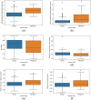
Illustration of box plots showing the discriminatory performance of some prominent vessel features extracted with the proposed approach. (a) Neighboring temperature increase, (b) contralateral temperature increase, (c) area ratio, (d) mirror overlap, (e) relative increase in the number of branches between the breasts, and (f) difference of vessel extent between the breasts.
Fig. 5
Fig. 5
Illustration of box plots showing lower discriminatory performance of skeleton features when we consider their absolute values instead of the proposed relative values. (a) Absolute number of branches and (b) absolute vessel extent.

References

    1. Sung H., et al. , “Global cancer statistics 2020: GLOBOCAN estimates of incidence and mortality worldwide for 36 cancers in 185 countries,” CA: Cancer J. Clin. 71(3), 209–249 (2021).10.3322/caac.21660 - DOI - PubMed
    1. Merino Bonilla J. A., Torres Tabanera M., Ros Mendoza L. H., “Breast cancer in the 21st century: from early detection to new therapies,” Radiología 59(5), 368–379 (2017).10.1016/j.rx.2017.06.003 - DOI - PubMed
    1. Smith R. A., et al. , “Breast cancer in limited-resource countries: early detection and access to care,” Breast J. 12(s1), S16–S26 (2006).BRJOFK10.1111/j.1075-122X.2006.00200.x - DOI - PubMed
    1. Coleman C., “Early detection and screening for breast cancer,” Semin. Oncol. Nurs. 33(2), 141–155 (2017).10.1016/j.soncn.2017.02.009 - DOI - PubMed
    1. Folkman J., “What is the evidence that tumors are angiogenesis dependent?” J. Natl. Cancer Inst. 82(1), 4–6 (1990).JNCIEQ10.1093/jnci/82.1.4 - DOI - PubMed