

The Science of Anastomotic Healing
- PMID: 35937614
- PMCID: PMC9355065
- DOI: 10.1016/j.scrs.2022.100879
The Science of Anastomotic Healing
Abstract
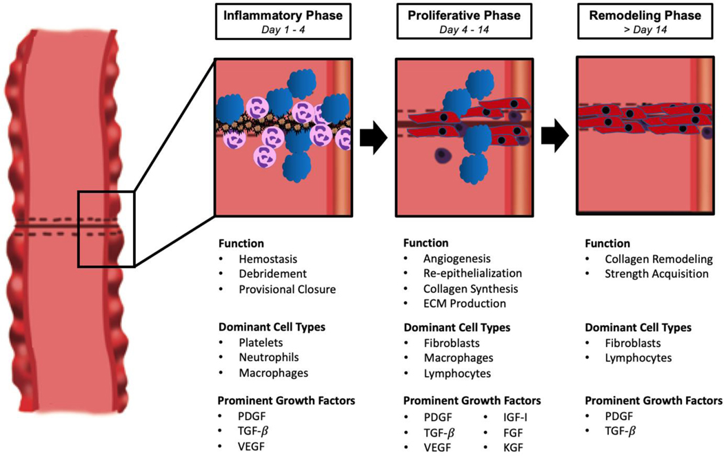
Intestinal anastomotic tissue follows a similar pattern of healing that is seen in all tissues with characteristic inflammatory, proliferative, and remodeling phases. Several aspects of intestinal healing are distinct from other tissues, however, including its time course and interaction with the environment of the gastrointestinal tract. As the anastomosis progresses through each stage, initial inflammatory cells are replaced by collagen-producing fibroblasts that generate the anastomosis' strength. A complex network of cell-to-cell signaling mediates this process through the release of cytokines and growth factors including platelet-derived growth factor (PDGF), transforming growth factor-β (TGF-β), and vascular endothelial growth factor (VEGF). Interventions based on these signaling pathways have been shown to improve anastomotic strength in animals, though methods for improving anastomotic healing in human patients remain unclear. Given the risks associated with anastomotic failure in patients, there is value in monitoring inflammatory markers and cytokines that can indicate the presence of a leak.
Keywords: Anastomotic healing; anastomotic leak; collagen remodeling; growth factors.
Conflict of interest statement
Disclosure: The author reports no potential conflicts.
Figures
References
-
- Thompson SK, Chang EY, Jobe BA. Clinical review: Healing in gastrointestinal anastomoses, part I. Microsurgery. 2006;26:131–136. - PubMed
-
- Nurden AT, Nurden P, Sanchez M, Andia I, Anitua E. Platelets and wound healing. Front Biosci. 2008;13:3532–3548. - PubMed
-
- Witte MB, Barbul A. General principles of wound healing. Surg Clin North Am. 1997;77:509–528. - PubMed
-
- Li J, Chen J, Kirsner R. Pathophysiology of acute wound healing. Clin Dermatol. 2007;25:9–18. - PubMed
Grants and funding
LinkOut - more resources
Full Text Sources
Research Materials