. 2022 Jul 22:2022:1422185.
doi: 10.1155/2022/1422185. eCollection 2022.

Nanoscale Features of Gambogic Acid Induced ROS-Dependent Apoptosis in Esophageal Cancer Cells Imaged by Atomic Force Microscopy

Affiliations

Nanoscale Features of Gambogic Acid Induced ROS-Dependent Apoptosis in Esophageal Cancer Cells Imaged by Atomic Force Microscopy

Jianxin Liu et al. Scanning. .

Abstract

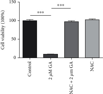
Gambogic acid (GA), a kind of polyprenylated xanthone derived from Garcinia hanburyi tree, has showed spectrum anticancer effects both in vitro and in vivo with low toxicity. However, up to now, there is little information about the effects of GA on esophageal cancer. In this study, we aim to test the anticancer effects of GA on esophageal cancer EC9706 cells. We established a nanoscale imaging method based on AFM to evaluate the reactive oxygen species- (ROS-) mediated anticancer effects of GA on esophageal cancer regarding the morphological and ultrastructural changes of esophageal cancer cells. The obtained results demonstrated that GA could inhibit cell proliferation, induce apoptosis, induce cell cycle arrest, and induce mitochondria membrane potential disruption in a ROS-dependent way. And using AFM imaging, we also found that GA could induce the damage of cellular morphology and increase of membrane height distribution and membrane roughness in EC9706 cells, which could be reversed by the removal of GA-induced excessive intracellular ROS. Our results not only demonstrated the anticancer effects of GA on EC9706 cells in ROS-dependent mechanism but also strongly suggested AFM as a powerful tool for the detection of ROS-mediated cancer cell apoptosis on the basis of imaging.


Conflict of interest statement

The authors declare no conflict of interest.

Figures

Figure 1
Figure 1
Effects of gambogic acid on the viability of EC9706 cells after 24 h treatment. n = 3; p < 0.05,  ∗∗p < 0.01, and∗∗∗p < 0.001.
Figure 2
Figure 2
Effects of gambogic acid on ROS production in EC9706 cells after 3 h treatment. Intracellular ROS level of (a) control, (b) 0.4 μM gambogic acid-treated, (c) 1.2 μM gambogic acid-treated, (d) 2 μM gambogic acid-treated, (e) 5 mM NAC+2 μM gambogic acid-treated, and (f) 5 mM NAC-treated EC9706 cells. EC9706 cells in (e, f) were pretreated with 5 mM NAC for 1 h and then treated with or without gambogic acid for 3 h to determine the effects of ROS scavenger-NAC on gambogic acid-induced ROS production in EC9706 cells.
Figure 3
Figure 3
Effects of ROS scavenger-NAC on gambogic acid inhibited viability of EC9706 cells. EC9706 cells were pretreated with 5 mM NAC for 1 h and then treated with or without gambogic acid for 24 h. n = 3; ∗∗∗p < 0.001.
Figure 4
Figure 4
Effects of gambogic acid on apoptosis of EC9706 cells after 24 h treatment. Intracellular ROS level of (a) control, (b) 0.4 μM gambogic acid-treated, (c) 1.2 μM gambogic acid-treated, (d) 2 μM gambogic acid-treated, (e) 5 mM NAC+2 μM gambogic acid-treated, and (f) 5 mM NAC-treated EC9706 cells. EC9706 cells in (e, f) were pretreated with 5 mM NAC for 1 h and then treated with or without gambogic acid for 24 h to determine the effects of ROS scavenger-NAC on gambogic acid induced apoptosis in EC9706 cells.
Figure 5
Figure 5
Effects of gambogic acid on cell cycle distribution of EC9706 cells after 24 h treatment. Intracellular ROS level of (a) control, (b) 0.4 μM gambogic acid-treated, (c) 1.2 μM gambogic acid-treated, (d) 2 μM gambogic acid-treated, (e) 5 mM NAC+2 μM gambogic acid-treated, and (f) 5 mM NAC-treated EC9706 cells. EC9706 cells in (e, f) were pretreated with 5 mM NAC for 1 h and then treated with or without gambogic acid for 24 h to determine the effects of ROS scavenger-NAC on gambogic acid induced cell cycle arrest in EC9706 cells.
Figure 6
Figure 6
Effects of gambogic acid on mitochondrial membrane potential of EC9706 cells after 24 h treatment. Intracellular ROS level of (a) control, (b) 0.4 μM gambogic acid-treated, (c) 1.2 μM gambogic acid-treated, (d) 2 μM gambogic acid-treated, (e) 5 mM NAC+2 μM gambogic acid-treated, and (f) 5 mM NAC-treated EC9706 cells. EC9706 cells in (e, f) were pretreated with 5 mM NAC for 1 h and then treated with or without gambogic acid for 24 h to determine the effects of ROS scavenger-NAC on gambogic acid induced mitochondrial membrane potential disruption in EC9706 cells.
Figure 7
Figure 7
Effects of gambogic acid on cell morphology of EC9706 cells after 24 h treatment. AFM morphology imaging of (a) control, (b) 0.4 μM gambogic acid-treated, (c) 1.2 μM gambogic acid-treated, (d) 2 μM gambogic acid-treated, (e) 5 mM NAC+2 μM gambogic acid-treated, and (f) 5 mM NAC-treated EC9706 cells. (A1-F1) Topography images and (A2-F2) their corresponding deflection error images. (A3-F3) Enlarged topography images as indicated by the blue frames in (A1-F1) and (A4-F4) their corresponding deflection error images. Scale bar: 20 μm. EC9706 cells in (e, f) were pretreated with 5 mM NAC for 1 h and then treated with or without gambogic acid for 24 h to determine the effects of ROS scavenger-NAC on gambogic acid induced morphological damage in EC9706 cells by AFM imaging in air with contact mode.
Figure 8
Figure 8
Effects of gambogic acid on cell membrane ultrastructure of EC9706 cells after 24 h treatment. AFM morphology and membrane ultrastructure images of (a) control, (b) 0.4 μM gambogic acid-treated, (c) 1.2 μM gambogic acid-treated, (d) 2 μM gambogic acid-treated, (e) 5 mM NAC+2 μM gambogic acid-treated, and (f) 5 mM NAC-treated EC9706 cells. (A1-F1) Topography images of EC9706 cells, scale bar: 10 μm. (A2-F2) Enlarged cell membrane ultrastructure images of EC9706 cells as indicated by the blue frames in (A1-F1); scale bar: 400 nm. EC9706 cells in (e, f) were pretreated with 5 mM NAC for 1 h and then treated with or without gambogic acid for 24 h to determine the effects of ROS scavenger-NAC on gambogic acid induced membrane ultrastructure changes in EC9706 cells by AFM imaging in air with contact mode.
Figure 9
Figure 9
Statistical analysis of the effects of gambogic acid on membrane ultrastructure of EC9706 cells after 24 h treatment and the effects of ROS scavenger-NAC on gambogic acid induced EC9706 cell membrane ultrastructural changes determined by AFM. (a) Root mean square roughness (Rq) and (b) average roughness (Ra) analyzed from 2 × 2 μm frame ultrastructure images of EC9706 cells. n = 10; ∗∗p < 0.01 and∗∗∗p < 0.001.

Similar articles

Cited by

References

    1. Kamangar F., Chow W. H., Abnet C. C., Dawsey S. M. Environmental causes of esophageal cancer. Gastroenterology Clinics of North America . 2009;38(1):27–57. doi: 10.1016/j.gtc.2009.01.004. - DOI - PMC - PubMed
    1. Zheng S., Vuitton L., Sheyhidin I., Vuitton D. A., Zhang Y., Lu X. Northwestern China: a place to learn more on oesophageal cancer. Part one: behavioural and environmental risk factors. European Journal of Gastroenterology & Hepatology . 2010;22(8):917–925. doi: 10.1097/MEG.0b013e3283313d8b. - DOI - PubMed
    1. Liesenklas W., Auterhoff H. Die Konstitution der Gambogasäure und ihre Isomerisierung 4. Mitt.: Chemie des Gummigutts. Archiv der Pharmazie und Berichte der Deutschen Pharmazeutischen Gesellschaft . 1966;299(9):797–798. doi: 10.1002/ardp.19662990911. - DOI - PubMed
    1. Wang J., Zhao L., Hu Y., et al. Studies on chemical structure modification and biology of a natural product, gambogic acid (I): synthesis and biological evaluation of oxidized analogues of gambogic acid. European Journal of Medicinal Chemistry . 2009;44(6):2611–2620. doi: 10.1016/j.ejmech.2008.09.034. - DOI - PubMed
    1. Huang H., Chen D., Li S., et al. Gambogic acid enhances proteasome inhibitor-induced anticancer activity. Cancer Letters . 2011;301(2):221–228. doi: 10.1016/j.canlet.2010.12.015. - DOI - PMC - PubMed