

doi: 10.4103/ojo.ojo_420_20.
eCollection 2022 May-Aug.
"Out of the ashes and into the fire"- Choroidal neovascular membrane at the intersection of traumatic choroidal rupture and brittle bruch's membrane
Affiliations
- PMID: 35937742
- PMCID: PMC9351943
- DOI: 10.4103/ojo.ojo_420_20
Item in Clipboard
"Out of the ashes and into the fire"- Choroidal neovascular membrane at the intersection of traumatic choroidal rupture and brittle bruch's membrane
Oman J Ophthalmol.
.
No abstract available
Keywords: Angioid streaks; choroidal neovascular membrane; trauma.
Conflict of interest statement
There are no conflicts of interest.
Figures
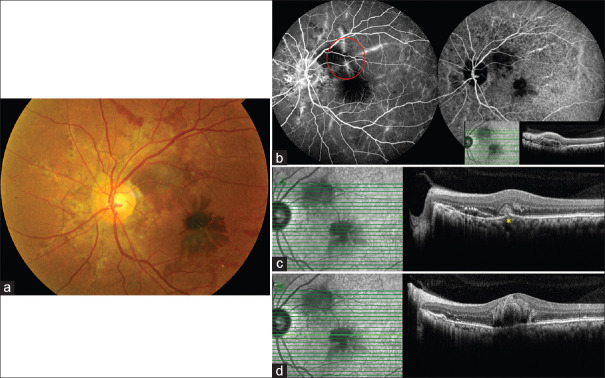
(a) Fundus photograph demonstrating the angioid streaks and peripapillary subretinal hemorrhage with a dark multilayered hemorrhage at the fovea, (b) FFA and ICG image showing the peripapillary ring and hyperfluorescent wavy lines denoting the angioid streaks, blocked fluoresence at the macula, and in the peripapillary region with faint lacy hyperfluoresence (denoted by red circle). Inset SD-OCT line scan through the intersection of choroidal rupture and angioid streak showing RPE elevation and subretinal hyperreflectivity and hyperreflective foci in the inner retinal layers, (c) SD-OCT superior to the fovea showing Bruch's membrane breach denoted by asterisk in yellow and subretinal hyperreflectivity, (d) SD-OCT through the fovea showing altered foveal contour, hyperreflectivity in multiple layers, and obscuration of deeper structures. FFA: Fundus fluorescein angiography, ICG: Indocyanine green, SD-OCT: Spectral domain optical coherence tomography, RPE: Retinal pigment epithelium
Similar articles
-
[Subretinal neovascularization in angioid streaks].Tunis Med. 2001 Mar;79(3):161-4. Tunis Med. 2001. PMID: 11471445 French.
-
Histologic and morphometric analysis of the choroid, Bruch's membrane, and retinal pigment epithelium in postmortem eyes with age-related macular degeneration and histologic examination of surgically excised choroidal neovascular membranes.Surv Ophthalmol. 1999 Oct;44 Suppl 1:S10-32. doi: 10.1016/s0039-6257(99)00086-7. Surv Ophthalmol. 1999. PMID: 10548114
-
[Subretinal neovascular membrane in angioid streaks treated with intravitreal bevacizumab].Arch Soc Esp Oftalmol. 2014 May;89(5):203-6. doi: 10.1016/j.oftal.2012.11.010. Epub 2013 Apr 3. Arch Soc Esp Oftalmol. 2014. PMID: 24269464 Spanish.
-
[Angioid streaks].J Fr Ophtalmol. 2012 Dec;35(10):838-45. doi: 10.1016/j.jfo.2012.05.003. Epub 2012 Oct 6. J Fr Ophtalmol. 2012. PMID: 23046745 Review. French.
-
Understanding angioid streaks.J Am Optom Assoc. 1997 May;68(5):309-24. J Am Optom Assoc. 1997. PMID: 9170798 Review.
References
LinkOut - more resources
Full Text Sources