Review
. 2022 Jun 29;15(2):133-139.
doi: 10.4103/ojo.ojo_215_21. eCollection 2022 May-Aug.

Management of neurovascular emergencies with ophthalmic manifestations

Affiliations
Review

Management of neurovascular emergencies with ophthalmic manifestations

Jawahar Lal Goyal et al. Oman J Ophthalmol. .

Abstract

Patients with neurovascular disorders sometimes approach the ophthalmologists with mild ophthalmic clinical features such as conjunctival congestion, slowly progressive proptosis, lateral rectus palsies and at other times with ophthalmic emergencies like sudden increase in proptosis, ophthalmoplegia, diplopia, and ptosis before the onset of neurological manifestations which may be life-threatening if not detected in time. The aim of this article is to focus on ophthalmic manifestations of neurovascular emergencies and role of ophthalmologists in its management. In this communication, to make the ophthalmologist aware of clinical presentations, the imaging modality of choice, diagnostic features, medical and interventional treatments. We have searched PubMed, Web of Science, Google Scholar and reviewed some of the commonly encountered neurovascular emergencies with ocular manifestations such as carotid-cavernous fistula, cerebral venous sinus thrombosis, cerebral artery aneurysms, arterio-venous malformations.

Keywords: Carotid-cavernous fistula; cavernous sinus thrombosis; cerebral aneurysm; neurovascular emergency; ophthalmic manifestations.


Conflict of interest statement

There are no conflicts of interest.

Figures
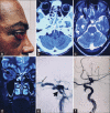
Figure 1
Figure 1
(a) Congestive changes with proptosis in carotid-cavernous fistula. (b) Computed tomography showing proptosis (denoted by thin white arrow) and hemosinus (denoted by thick white arrow). (c and d) T2 images showing dilated superior ophthalmic vein (denoted by thick white arrow in c and thin white arrow in d) and enlarged cavernous sinus (denoted by thin white arrow in c). (e) Digital subtraction angiography showing steal phenomenon, fistula site (denoted by thick black arrow) and dilated superior ophthalmic vein (denoted by thin black arrow) are seen. (f) Digital subtraction angiography showing fistula obliteration using balloon (denoted by thin white arrow)
Figure 2
Figure 2
(a) T1-weighted axial postcontrast images of brain showing filling defect in right transverse sinus suggestive of thrombosis (denoted by white arrow). Please note associated distension suggesting acute nature. (b) Contrast-enhanced magnetic resonance venography showing nonvisualization of right transverse sinus consistent with thrombosis
Figure 3
Figure 3
(a) Noncontrast Computed tomography showing partially thrombosed aneurysm from left cavernous internal carotid artery (denoted by white arrow). (b) T1 postcontrast image showing aneurysm. (c) Digital subtraction angiography image of left cavernous internal carotid artery showing aneurysm (denoted by white arrow). (d) Digital subtraction angiography image showing flow diverter device (denoted by white arrow) in left cavernous internal carotid artery reducing flow to aneurysm
Figure 4
Figure 4
(a) NCCT brain showing aneurysm at internal carotid artery bifurcation (denoted by blue arrow). (b) Computed tomography angiography showing aneurysm at internal carotid artery -Posterior communicating artery junction (denoted by blue arrow). (c) Magnetic resonance angiography showing aneurysm (denoted by blue arrow). (d) Postcontrast magnetic resonance imaging showing coil mass in aneurysm (denoted by blue arrow). (e) Digital subtraction angiography showing aneurysm (denoted by blue arrow). (f) Digital subtraction angiography (postcoiling) showing coil mass in aneurysm (denoted by blue arrow)
Figure 5
Figure 5
(a) NCCT brain showing lesion (denoted by white arrow) in the occipital lobe. (b) T1 image showing lesion (denoted by white arrow). (c) T2 image showing flow (denoted by white arrow) voids within lesion suggesting pial arteriovenous malformation. (d) Digital subtraction angiography image showing pial AVM. (e) Digital subtraction angiography image postembolization showing the onyx cast (denoted by black arrows). Note made of microcather (denoted by yellow arrow)

References

    1. Rahman WT, Griauzde J, Chaudhary N, Pandey AS, Gemmete JJ, Chong ST. Neurovascular emergencies: Imaging diagnosis and neurointerventional treatment. Emerg Radiol. 2017;24:183–93. - PubMed
    1. Barker WF, Stern WE, Krayenbuhl H, Senning A. Carotid endarterectomy complicated by carotid-cavernous sinus fistula. Ann Surg. 1968;167:568–72. - PMC - PubMed
    1. Henderson AD, Miller NR. Carotid-cavernous fistula: Current concepts in aetiology, investigation, and management. Eye (Lond) 2018;32:164–72. - PMC - PubMed
    1. Zhang Y, Zheng H, Zhou M, He L. Teaching neuroimages: Carotid-cavernous fistula caused by fibromuscular dysplasia. Neurology. 2014;82:e134–5. - PubMed
    1. Srinivas HV, Murthy S, Brown R. Is Valsalva manoeuvre useful in diagnosing dural caroticocavernous fistulas? Eye (Lond) 2005;19:1226–7. - PubMed