Review
. 2022 Feb 2;6(4):161-172.
doi: 10.1515/iss-2021-0028. eCollection 2021 Dec 1.

Image-guided surgery and novel intraoperative devices for enhanced visualisation in general and paediatric surgery: a review

Affiliations
Review

Image-guided surgery and novel intraoperative devices for enhanced visualisation in general and paediatric surgery: a review

Laura Privitera et al. Innov Surg Sci. .

Abstract

Fluorescence guided surgery, augmented reality, and intra-operative imaging devices are rapidly pervading the field of surgical interventions, equipping the surgeon with powerful tools capable of enhancing the surgical visualisation of anatomical normal and pathological structures. There is a wide range of possibilities in the adult population to use these novel technologies and devices in the guidance for surgical procedures and minimally invasive surgeries. Their applications and their use have also been increasingly growing in the field of paediatric surgery, where the detailed visualisation of small anatomical structures could reduce procedure time, minimising surgical complications and ultimately improve the outcome of surgery. This review aims to illustrate the mechanisms underlying these innovations and their main applications in the clinical setting.

Keywords: augmented reality; fluorescence-guided surgery; general surgery; image-guided surgery; intra-operative visualisation; novel devices; optical imaging; paediatric surgery.


Conflict of interest statement

Competing interests: Authors state no conflict of interest.

Figures
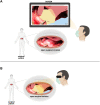
Figure 1:
Figure 1:
AR in the surgical field through the visualisation of data projected on a screen (panel A) or with a head-mounted display (black arrow in panel B) that superimposed objects onto real-time images (panel B). The picture shows a tumour (yellow) before surgical resection. The area in orange shows the extension of the tumour into the liver (red). The bowel is marked in light blue.
Figure 2:
Figure 2:
Schematic representation of contrast-enhanced ultrasound mechanism of action, with intravenously administered contrast agent. Ultrasound contrast agents consist of gas-filled microbubbles (1–10 µm) with a lipid, protein, or polymer shell. The pressure changes induced by the ultrasonic waves lead microbubbles to contract (compression) and expand (rarefaction) to a higher degree compared to the surrounding tissues. This, along with the impedance mismatch between the microbubble and surrounding fluid caused by the gas, makes the bubbles highly echogenic. Abbreviations: RBC, red blood cell; EC, endothelial cell.
Figure 3:
Figure 3:
Schematic representation of photoacoustic imaging mechanism of action. The absorption of light by endogenous chromophores (or pigments) generates heat, leading to a pressure change. The resulting fleeting expansions generate an ultrasound wave which can then be detected and used to produce clear, high-resolution images of biological structures.

Similar articles

Cited by

References

    1. Paraboschi I, De Coppi P, Stoyanov D, Anderson J, Giuliani S. Fluorescence imaging in pediatric surgery: state-of-the-art and future perspectives. J Pediatr Surg. 2020 Aug 15 doi: 10.1016/j.jpedsurg.2020.08.004. Epub ahead of print. - DOI - PubMed
    1. Guha D, Alotaibi NM, Nguyen N, Gupta S, McFaul C, Yang VXD. Augmented reality in neurosurgery: a review of current concepts and emerging applications. Can J Neurol Sci. 2017;44:235–45. doi: 10.1017/cjn.2016.443. - DOI - PubMed
    1. Paul P, Fleig O, Jannin P. Augmented virtuality based on stereoscopic reconstruction in multimodal image-guided neurosurgery: methods and performance evaluation. IEEE Trans Med Imag. 2005;24:1500–11. doi: 10.1109/TMI.2005.857029. - DOI - PubMed
    1. Giordano M, Samii A, Lawson McLean AC, Bertalanffy H, Fahlbusch R, Samii M, et al. Intraoperative magnetic resonance imaging in pediatric neurosurgery: safety and utility. J Neurosurg Pediatr. 2017;19:77–84. doi: 10.3171/2016.8.PEDS15708. - DOI - PubMed
    1. Di Carlo HN, Maruf M, Massanyi EZ, Shah B, Tekes A, Gearhart JP. 3-dimensional magnetic resonance imaging guided pelvic floor dissection for bladder exstrophy: a single arm trial. J Urol. 2019;202:406–12. doi: 10.1097/JU.0000000000000210. - DOI - PubMed