

Cerebral blood flow network differences correlated with cognitive impairment in mild traumatic brain injury
- PMID: 35937870
- PMCID: PMC9355478
- DOI: 10.3389/fnins.2022.969971
Cerebral blood flow network differences correlated with cognitive impairment in mild traumatic brain injury
Abstract
Purpose: To examine whether the cerebral blood flow (CBF) and CBF connectivity differences are sex-specific and whether these differences are correlated with cognitive impairment in mTBI.
Methods: Resting-state perfusion magnetic resonance imaging was performed in 40 patients with acute mTBI and 40 healthy controls by using pseudocontinuous arterial spin labeling within 14 days following injury. The differences in normalized CBF were first compared and CBF connectivity of the brain regions with significant CBF differences were compared next. The association between the normalized CBF and CBF connectivity differences and cognitive function were further investigated.
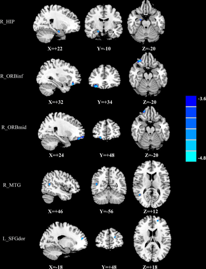
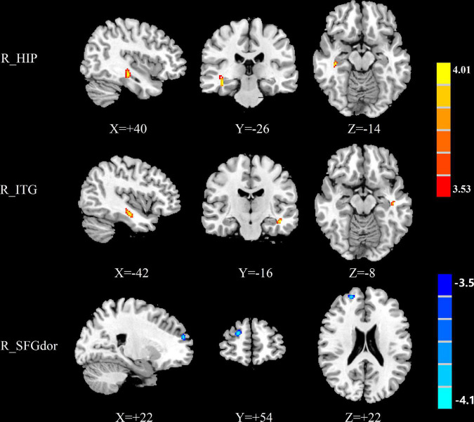
Results: Men patients had lower normalized CBF in the frontal gyrus, temporal gyrus and hippocampus and decreased negative CBF connectivity between brain regions including the hippocampus, temporal gyrus, postcentral gyrus and lenticular nucleus, putamen, compared with men controls. Women patients had lower normalized CBF in the frontal gyrus, however had higher normalized CBF in the temporal gyrus and hippocampus, compared with women controls. Additionally, women patients showed increased positive CBF connectivity between the seed region of interest (ROI) of the right inferior temporal gyrus and temporal gyrus and frontal gyrus, and had increased positive CBF connectivity between the seed ROI of the right hippocampus and the temporal gyrus. Furthermore, men patients had higher CBF in the right middle temporal gyrus and left precentral gyrus than women patients.
Conclusion: This study provides evidence of sex differences in both decreased and increased CBF and CBF connectivity and association with cognitive outcome in the acute stage after mTBI.
Keywords: arterial spin labeling; cerebral blood flow; cognitive impairment; magnetic resonance imaging; mild traumatic brain injury.
Copyright © 2022 Duan, Liu, Li, Lu and Chen.
Conflict of interest statement
The authors declare that the research was conducted in the absence of any commercial or financial relationships that could be construed as a potential conflict of interest. The handling editor VM and reviewer ZX declared a past co-authorship with the authors Y-CC, LL, and FL.
Figures


Similar articles
-
Cerebral Blood Flow and Its Connectivity Deficits in Mild Traumatic Brain Injury at the Acute Stage.Neural Plast. 2020 Jul 1;2020:2174371. doi: 10.1155/2020/2174371. eCollection 2020. Neural Plast. 2020. PMID: 32684919 Free PMC article.
-
Arterial spin labeling magnetic resonance evaluates changes of cerebral blood flow in patients with mild traumatic brain injury.Zhong Nan Da Xue Xue Bao Yi Xue Ban. 2022 Aug 28;47(8):1016-1024. doi: 10.11817/j.issn.1672-7347.2022.210754. Zhong Nan Da Xue Xue Bao Yi Xue Ban. 2022. PMID: 36097769 Free PMC article. Chinese, English.
-
Functional connectivity dysfunction of insular subdivisions in cognitive impairment after acute mild traumatic brain injury.Brain Imaging Behav. 2020 Jun;14(3):941-948. doi: 10.1007/s11682-020-00288-5. Brain Imaging Behav. 2020. PMID: 32304021 Free PMC article.
-
Alterations of Cerebral Blood Flow and its Connectivity Patterns Measured with Arterial Spin Labeling in Mild Cognitive Impairment.Curr Alzheimer Res. 2023;20(8):567-576. doi: 10.2174/0115672050241163231017073139. Curr Alzheimer Res. 2023. PMID: 37921165
-
Aberrant pattern of regional cerebral blood flow in mild cognitive impairment: A meta-analysis of arterial spin labeling magnetic resonance imaging.Front Aging Neurosci. 2022 Sep 1;14:961344. doi: 10.3389/fnagi.2022.961344. eCollection 2022. Front Aging Neurosci. 2022. PMID: 36118708 Free PMC article.
Cited by
-
Cognitive impairment in diffuse axonal injury patients with favorable outcome.Front Neurosci. 2023 Jan 25;17:1077858. doi: 10.3389/fnins.2023.1077858. eCollection 2023. Front Neurosci. 2023. PMID: 36761409 Free PMC article.
-
Disrupted resting-state functional connectivity and network topology in mild traumatic brain injury: an arterial spin labelling study.Brain Commun. 2023 Sep 30;5(5):fcad254. doi: 10.1093/braincomms/fcad254. eCollection 2023. Brain Commun. 2023. PMID: 37829696 Free PMC article.
-
Resting-State Brain Activity Changes and Their Genetic Correlates in Mild Traumatic Brain Injury.Hum Brain Mapp. 2025 Jun 1;46(8):e70259. doi: 10.1002/hbm.70259. Hum Brain Mapp. 2025. PMID: 40509941 Free PMC article.
-
From spreading depolarization to blood-brain barrier dysfunction: navigating traumatic brain injury for novel diagnosis and therapy.Nat Rev Neurol. 2024 Jul;20(7):408-425. doi: 10.1038/s41582-024-00973-9. Epub 2024 Jun 17. Nat Rev Neurol. 2024. PMID: 38886512 Review.
References
-
- Cui L. B., Wang L. X., Tian P., Wang H. N., Cai M., Guo F., et al. (2017). Aberrant perfusion and its connectivity within default mode network of first-episode drug-naïve schizophrenia patients and their unaffected first-degree relatives. Sci. Rep. 7:16201. 10.1038/s41598-017-14343-7 - DOI - PMC - PubMed
LinkOut - more resources
Full Text Sources