. 2022 Jul 13;25(8):104754.
doi: 10.1016/j.isci.2022.104754. eCollection 2022 Aug 19.

The endoplasmic reticulum membrane complex promotes proteostasis of GABAA receptors

Affiliations

The endoplasmic reticulum membrane complex promotes proteostasis of GABAA receptors

Angela L Whittsette et al. iScience. .

Abstract

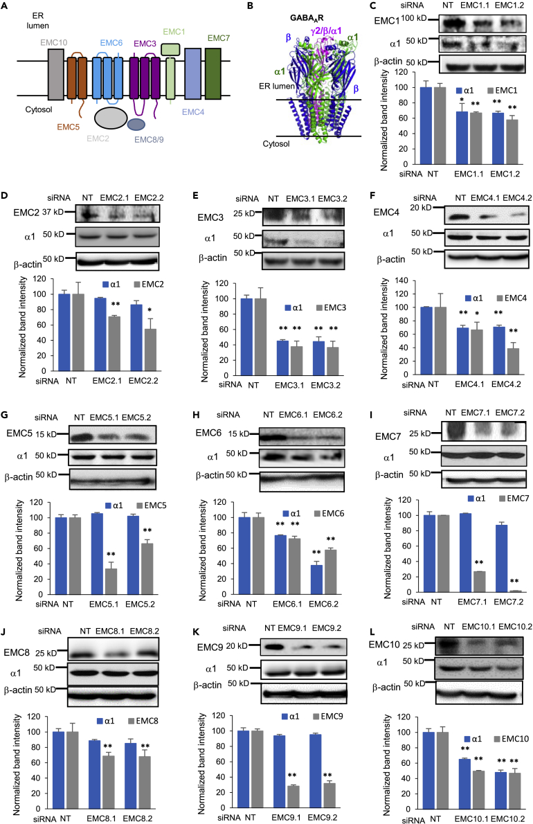
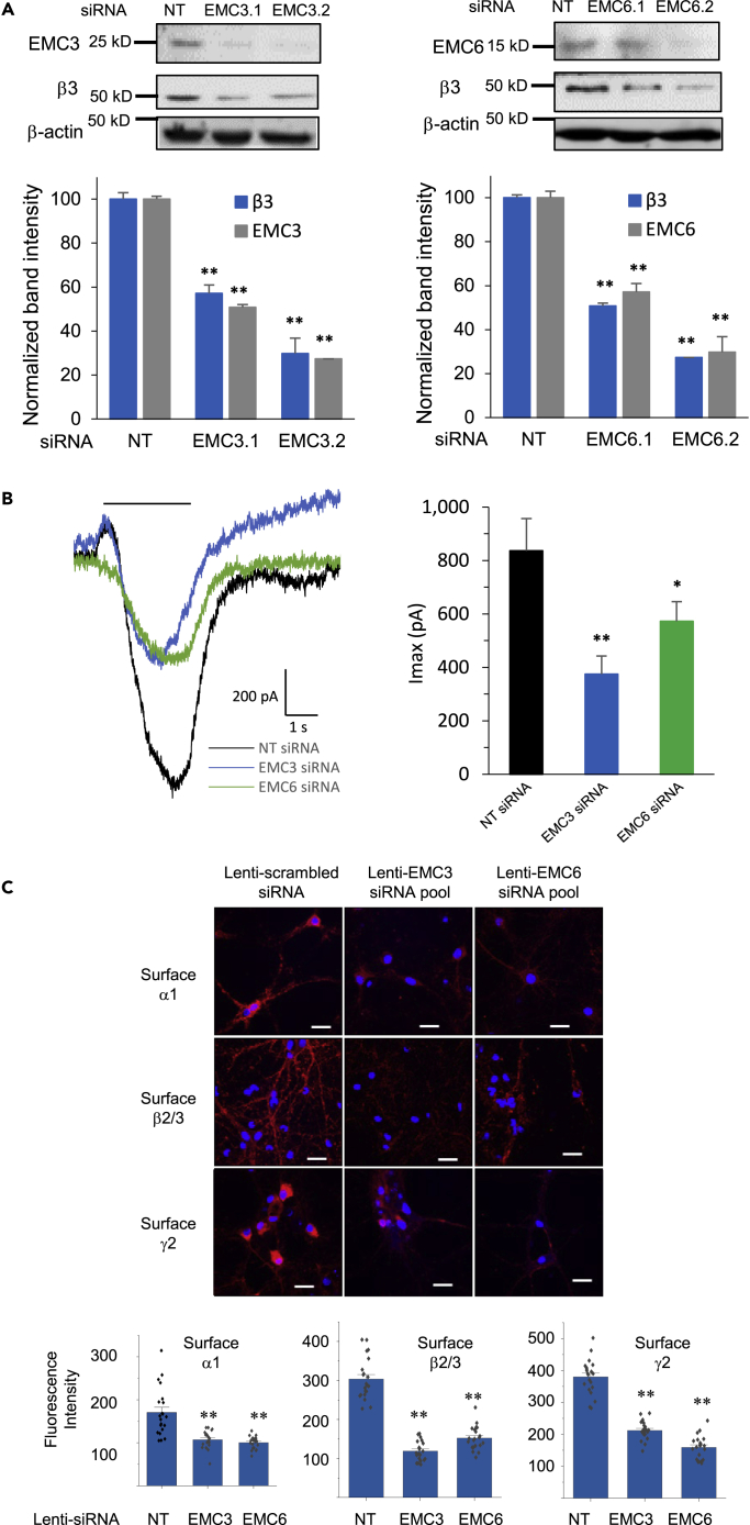
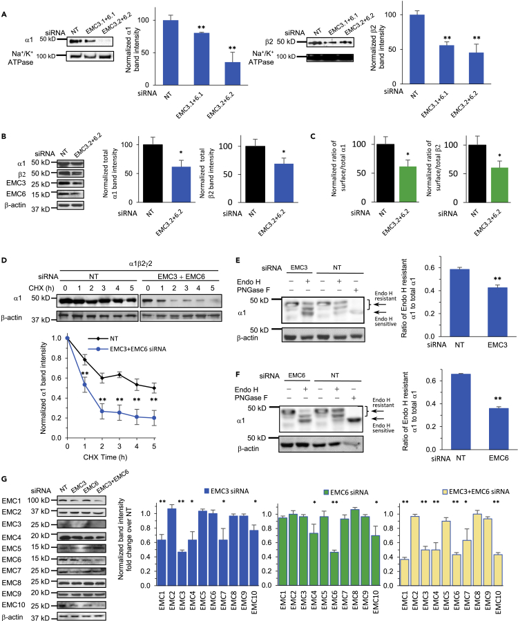
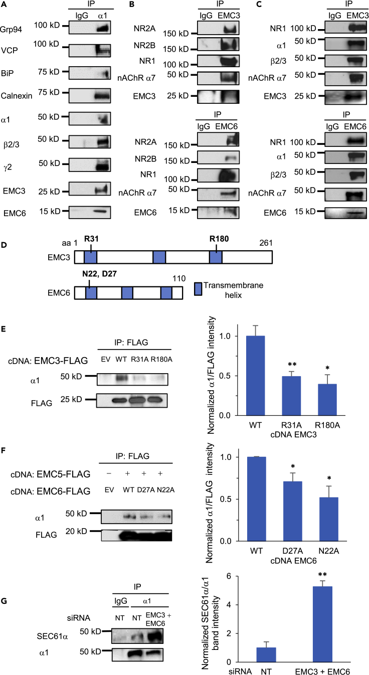
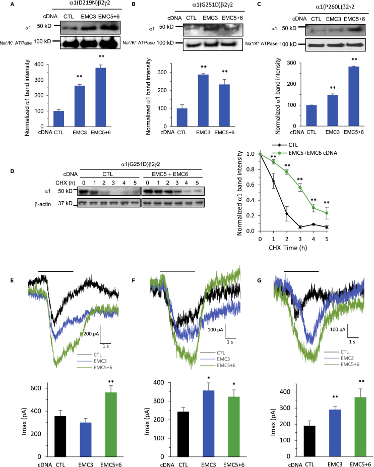
The endoplasmic reticulum membrane complex (EMC) plays a critical role in the biogenesis of tail-anchored proteins and a subset of multi-pass membrane proteins in the endoplasmic reticulum (ER). However, because of nearly exclusive expression of neurotransmitter-gated ion channels in the central nervous system (CNS), the role of the EMC in their biogenesis is not well understood. In this study, we demonstrated that the EMC positively regulates the surface trafficking and thus function of endogenous γ-aminobutyric acid type A (GABAA) receptors, the primary inhibitory ion channels in the mammalian brain. Moreover, among ten EMC subunits, EMC3 and EMC6 have the most prominent effect, and overexpression of EMC3 or EMC6 is sufficient to restore the function of epilepsy-associated GABAA receptor variants. In addition, EMC3 and EMC6 demonstrate endogenous interactions with major neuroreceptors, which depends on their transmembrane domains, suggesting a general role of the EMC in the biogenesis of neuroreceptors.

Keywords: Biological sciences; Cell biology; Molecular biology; Molecular neuroscience; Neuroscience.


Conflict of interest statement

The authors declare no competing interests.

Figures

None
Graphical abstract
Figure 1
Figure 1
Effect of depleting individual EMC subunits on endogenous GABAA receptor α1 subunit protein levels (A) Schematics of ten EMC subunits. (B) Cartoon Representation of heteropentameric GABAA receptors. The most common subtype in the mammalian CNS consists of α1, β2/β3, and γ2 subunits. (C to L) Endogenous total GABAA receptor α1 subunits protein level change on knocking down individual EMC subunits. Mouse hypothalamus GT1-7 neuronal cells were transfected with siRNAs against EMC1 to EMC10, respectively. Two distinct siRNAs targeting each of the ten EMC subunits, designated as EMCn.1 and EMCn.2 (n = 1 to 10), were used to minimize the potential off-target effects. Forty-eight hours after transfection, proteins were extracted and analyzed by western blotting. β-actin was used as the loading control. Normalized band intensity was shown below the images (n = 3). Each data point is presented as mean ± SEM ∗, p< 0.05; ∗∗, p< 0.01. NT: Non-targeting scrambled siRNA.
Figure 2
Figure 2
Effect of EMC3 and EMC6 on the protein levels and whole-cell patch-clamping currents of endogenous GABAAreceptors (A) Mouse GT1-7 neurons were incubated with siRNA against EMC3 or EMC6 for 48 h. Proteins were extracted and analyzed by western blotting; normalized band intensity was shown below the images (n = 3), with β-actin as the loading control. (B) Mouse GT1-7 neurons were incubated with siRNA against EMC3 or EMC6 for 48 h. Whole-cell patch-clamping was performed on the cells with the IonFlux Mercury 16 ensemble plates at a holding potential of −60 mV. GABA (1 mM) was applied for 4 s, as indicated by the horizontal bar above the currents. The peak currents (Imax) were acquired and analyzed by Fluxion Data Analyzer (n = 6 - 10). NT: Non-targeting scrambled siRNA; pA, picoampere. (C) Confocal microscopy imaging of primary rat cortical neurons demonstrated reduced surface expression of GABAA receptors after siRNA treatment of EMC3 and EMC6 through lentivirus transduction. Lentiviruses were generated from transiently transfected HEK293T cells with the following plasmids and collected after 60 h from the media passing through 0.45 μm filter: EMC3- or EMC6-set of four siRNA lentivectors, packaging and envelop plasmids. At day-in-vitro (DIV) 6 of the primary rat cortical neurons, lentivirus transduction was carried out at a multiplicity-of-infection (MOI) of 10. At DIV 12, neurons were stained for cell surface GABAA receptor α1 subunits (top row), β2/β3 subunits (middle row), and γ2 subunits (bottom row), colored in red. DAPI staining for the nucleus was colored in blue. Scale bar = 20 μm. Quantification of the fluorescence intensity by using ImageJ was shown on the bottom after background correction from 20–30 neurons. Each data point is presented as mean ± SEM ∗, p< 0.05; ∗∗, p< 0.01.
Figure 3
Figure 3
EMC3 and EMC6 promote anterograde trafficking of GABAA receptors (A and B) Significant reduction of cell surface and total α1 and β2 subunits of GABAA receptors was observed when both EMC3 and EMC6 were knocked down. We carried out siRNA transfection in HEK293T cells stably expressing α1β2γ2 GABAA receptors. To test the surface expression of GABAA receptors, biotinylation experiments were performed 48 h after siRNA transfection of both EMC3 and EMC6 (A). Surface proteins were enriched through biotin-neutravidin affinity purification, and western blot analysis was applied to detect surface α1 and β2 subunits. Na+/K+ ATPase served as loading control of cell surface proteins. To test the total protein expression of GABAA receptors, cells were lysed and total proteins were collected and subjected to SDS-PAGE and western blot analysis (B). β-actin was used as the total protein loading control. Normalized band intensity was shown on the right to the blots (n = 3). (C) The ratio of the surface/total subunits of GABAA receptors was quantified, as a measure of their surface trafficking efficiency. Data was taken from (A) and (B) for the calculation. (D) HEK293T cells stably expressing α1β2γ2 GABAA receptors were transfected with non-targeting siRNA or siRNAs against EMC3 and EMC6. 48 h after transfection, cycloheximide (CHX) (100 μg/mL), a potent protein synthesis inhibitor, was added to cell culture media for the indicated time. Cells were then harvested, and total proteins were subjected to SDS-PAGE and western blot analysis. Quantification of the α1 band intensity was plotted against the incubation time with CHX (n = 3). (E and F) EMC3 and EMC6 promote GABAA receptors’ trafficking from the ER to Golgi as demonstrated through endoglycosidase H (Endo H) digestion. EMC3 or EMC6 siRNA transfection was applied in HEK293T cells stably expressing α1β2γ2 GABAA receptors; 48 h after transfection, proteins were extracted, and subjected to Endo H digestion and western blot analysis. Endo H resistant bands (top two bands in lanes two and 4) represent proteins that have correctly folded in the ER, trafficked to Golgi and fully modified with the N-linked complex glycans, thus becoming resistant to Endo H. On the other hand, acting upon proteins remaining in ER, Endo H may remove the high mannose structure after the asparaginyl-N-acetyl-D-glucosamine on the α1 subunits, generating Endo H sensitive bands (bottom band in lanes two and 4). The Peptide-N-Glycosidase F (PNGase F) enzyme-treated samples served as a control for unglycosylated α1 subunits (lane 5). Quantification of the ratio of Endo H resistant/total α1 band intensity, as a measure of the trafficking efficiency of α1 subunits, was shown on the right (n = 3). (G) HEK293T cells stably expressing α1β2γ2 GABAA receptors were transfected with non-targeting siRNA or siRNAs against EMC3, EMC6, or both EMC3 and EMC6. 48 h after transfection, cells were harvested, and total proteins were subjected to SDS-PAGE and western blot analysis. Quantification of the normalized individual EMC subunit band intensity was shown on the right panels (n = 3). Each data point is presented as mean ± SEM ∗, p< 0.05; ∗∗, p< 0.01. NT: Non-targeting scrambled siRNA.
Figure 4
Figure 4
Interactions of EMC3 and EMC6 with neurotransmitter-gated ion channels (A) Co-immunoprecipitation (Co-IP) from primary rat cortical neurons demonstrated endogenous interactions between α1 subunits of GABAA receptors and EMC3, EMC6, and a number of α1-interacting chaperones (BiP and calnexin) and ERAD factors (Grp94 and VCP). Neurons were plated onto 10-cm dishes at a density of one million per dish. At DIV 12, proteins were extracted for Co-IP. IgG was used as a negative control during the immunoprecipitation. n = 3. (B) Co-IP from primary rat cortical neurons demonstrated endogenous interactions between EMC3/EMC6 and a number of ion channels, including N-methyl-D-aspartate receptors (NMDARs, including NR1, NR2A and NR2B subunits) and nicotinic acetylcholine receptors (nAChR α7 subunit). n = 3. (C) Co-IP from mouse cortical homogenates, which were prepared from C57BL/6J mice between 8 and 10 weeks of age, demonstrated endogenous interactions between EMC3/EMC6 and selected ion channels. n = 3. (D) Schematic of the primary sequence of EMC3 and EMC6. R31 and R180 in EMC3 and N22 and D27 in EMC6 were reported to influence the biogenesis of EMC-dependent client proteins. (E) Mutation of R31A or R180A in EMC3 significantly reduced the interaction of EMC3 with GABAA α1 subunits. The cDNAs of FLAG-tagged EMC3, either in the wild type (WT) form or carrying appropriate mutations of R31A or R180A, were transiently transfected in HEK293T cells stably expressing α1β2γ2 GABAA receptors. 48 h after transfection, proteins were extracted from cell lysates and incubated with anti-FLAG M2 magnetic beads. The immuno-purified eluents were separated through SDS-PAGE gel, and western blot analysis was performed to detect α1 subunits and FLAG. Quantification of the band intensity of α1 over FLAG after immunoprecipitation was shown on the right (n = 3). (F) Mutation of D27A or N22A in EMC6 significantly reduced the interaction of EMC6 with GABAAR α1 subunits. Transfection of cDNAs was applied similarly as in E, however with co-application of FLAG-tagged EMC5 and EMC6 variants in HEK293T cells stably expressing α1β2γ2 GABAA receptors. Co-IP and visualization of protein bands were carried out the same way as in E as well. Quantification of the band intensity of α1 over FLAG-tagged EMC6 after immunoprecipitation was shown on the right (n = 3). (G) Significant increase of the interaction of SEC61α and α1 subunits of GABAA receptors was observed when both EMC3 and EMC6 were knocked down. We carried out siRNA transfection in HEK293T cells stably expressing α1β2γ2 GABAA receptors; 48 h after transfection, proteins were extracted from cell lysates and incubated with anti-α1 antibody. The immuno-purified eluents were separated through SDS-PAGE gel, and western blot analysis was performed to detect SEC61α and α1 subunits. Quantification of the band intensity of SEC61α over α1 after immunoprecipitation was shown on the right (n = 3). Each data point is presented as mean ± SEM ∗, p< 0.05; ∗∗, p< 0.01. NT: Non-targeting scrambled siRNA; IP: immunoprecipitation; EV: empty vector; WT: wild type.
Figure 5
Figure 5
Overexpression of EMC3, and EMC5 and EMC6 restores surface expression and whole-cell currents of disease-associated variants of GABAA receptors (A–C). Overexpression of EMC3 and EMC5/6 increased surface expression of α1 subunits of GABAAR in HEK293T cells stably expressing α1(D219N)β2γ2 (A), α1(G251D)β2γ2 (B) and α1(P260L)β2γ2 (C). We carried out cDNA transfection of EMC3, or co-application of EMC5 and EMC6, in corresponding HEK293T cells; 48 h after transfection, surface proteins were enriched through biotin-neutravidin affinity purification, and western blot analysis was applied to detect α1 subunits. Na+/K+ ATPase served as loading control of cell surface proteins. Normalized surface α1 band intensity was shown below the images (n = 3). (D) HEK293T cells stably expressing α1(G251D)β2γ2 GABAA receptors were transfected with empty vector control (CTL) or EMC5 and EMC6 cDNAs. 48 h after transfection, cycloheximide (CHX) (100 μg/mL), a potent protein synthesis inhibitor, was added to cell culture media for the indicated time. Cells were then harvested, and total proteins were subjected to SDS-PAGE and western blot analysis. Quantification of the α1 band intensity was plotted against the incubation time with CHX (n = 3). (E–G) Increased whole-cell patch-clamping currents of GABAA receptors were recorded in HEK293T cells stably expressing α1(D219N)β2γ2 (E), α1(G251D)β2γ2 (F) and α1(P260L)β2γ2 (G). Transfection of cDNA was applied the same way as in (A–C); 48 h after transfection, patch clamping was performed on the cells with the IonFlux Mercury 16 ensemble plates at a holding potential of −60 mV. GABA (100 μM) was applied for 4 s, as indicated by the horizontal bar above the currents. The peak currents (Imax) were acquired and analyzed by Fluxion Data Analyzer (n = 6 - 10). Each data point is presented as mean ± SEM ∗, p< 0.05; ∗∗, p< 0.01. CTL: Empty vector control sample.
Figure 6
Figure 6
Working model of the role of the EMC on the biogenesis of GABAA receptors in the ER The EMC facilitates the insertion of the transmembrane domains of GABAA receptor subunits into the lipid bilayer. ER chaperones, such as BiP and calnexin, promote the folding of the ER luminal domains of GABAA receptors for subsequent assembly into pentameric receptors in the ER.

Similar articles

Cited by

References

    1. Absalom N.L., Liao V.W.Y., Johannesen K.M.H., Gardella E., Jacobs J., Lesca G., Gokce-Samar Z., Arzimanoglou A., Zeidler S., Striano P., et al. Gain-of-function and loss-of-function GABRB3 variants lead to distinct clinical phenotypes in patients with developmental and epileptic encephalopathies. Nat. Commun. 2022;13:1822. doi: 10.1038/s41467-022-29280-x. - DOI - PMC - PubMed
    1. Akerman C.J., Cline H.T. Refining the roles of GABAergic signaling during neural circuit formation. Trends Neurosci. 2007;30:382–389. doi: 10.1016/j.tins.2007.06.002. - DOI - PubMed
    1. Bai L., You Q., Feng X., Kovach A., Li H. Structure of the ER membrane complex, a transmembrane-domain insertase. Nature. 2020;584:475–478. - PMC - PubMed
    1. Barnes E.M. Assembly and intracellular trafficking of GABA(A) receptors. Int. Rev. Neurobiol. 2001;48:1–29. - PubMed
    1. Cao X., An J., Cao Y., Lv J., Wang J., Ding Y., Lin X., Zhou X. EMC3 is essential for retinal organization and neurogenesis during mouse retinal development. Invest. Ophthalmol Vis. Sci. 2021;62:31. doi: 10.1167/iovs.62.2.31. - DOI - PMC - PubMed

LinkOut - more resources