

Consecutive central and branch retinal vein occlusions in the same eye of a young healthy COVID-19 patient: A unique case report
- PMID: 35938144
- PMCID: PMC9339164
- DOI: 10.1016/j.ajoc.2022.101669
Consecutive central and branch retinal vein occlusions in the same eye of a young healthy COVID-19 patient: A unique case report
Abstract
Purpose: To report a case of consecutive central retinal vein occlusion (CRVO) and branch retinal vein occlusion (BRVO) in the same eye correlated with coronavirus disease (COVID-19) of the otherwise healthy patient.
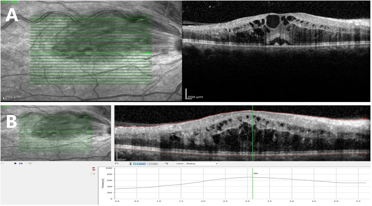
Observations: A 39-year-old woman with the diagnosis of COVID-19 infection for two weeks presented with a nonischemic central retinal vein occlusion (CRVO) in her right eye. The patient was on low-dose aspirin for anticoagulant prophylaxis (100 mg/day) for a week when the CRVO occurred. She had no history of any systemic risk factors for retinal vein occlusion (RVO) and her systemic evaluation failed to identify an etiology for her unilateral CRVO. While she was on monthly follow-up with no additional treatment, she experienced sudden visual acuity decrease in the same eye four months after the first RVO incident and one month after the cessation of aspirin intake. Her best corrected visual acuity (BCVA) was decreased from 20/25+ to 20/63. Her fundoscopic examination revealed increased intraretinal hemorrhages, dilated tortuous veins in the upper hemifield and macular edema. The central macular thickness measurement by optic coherence tomography was increased from 234 μm to 700 μm. The patient refused to undergo a fundus fluorescein angiography. After the diagnosis of the branch retinal vein occlusion with cystoid macular edema was done, the aspirin prophylaxis was restarted, and she received three intravitreal antivascular endothelial growth factor one month apart for her macular edema. Her BCVA improved to 20/20, and macular edema disappeared without any recurrence during the 6-month follow-up.
Conclusions and importance: To the best of our knowledge, this unique case is the first report of consecutive RVOs in the same eye of a healthy young patient associated with COVID-19. As our case report demonstrated, close follow-up and timely initiation of appropriate treatment could give rise to complete resolution of RVO.
Keywords: Anti-vascular endothelial growth factor; Branch retinal vein occlusion; COVID-19; Central retinal vein occlusion; Macular edema.
© 2022 Published by Elsevier Inc.
Conflict of interest statement
The following authors have no financial disclosures: AGA, None; EE, None.
Figures


Similar articles
-
Comparison of intravitreal bevacizumab upload followed by a dexamethasone implant versus dexamethasone implant monotherapy for retinal vein occlusion with macular edema.Ophthalmologica. 2012;228(2):110-6. doi: 10.1159/000338732. Epub 2012 Jun 23. Ophthalmologica. 2012. PMID: 22739239
-
"Off-label" use of intravitreal bevacizumab in non-ischemic macular edema secondary to retinal vein obstructions.Rom J Ophthalmol. 2016 Apr-Jun;60(2):90-95. Rom J Ophthalmol. 2016. PMID: 29450329 Free PMC article.
-
Case Report: Branch Retinal Vein Occlusion Post-mRNA SARS-CoV-2 (COVID-19) Vaccination.Optom Vis Sci. 2023 Nov 1;100(11):799-803. doi: 10.1097/OPX.0000000000002075. Optom Vis Sci. 2023. PMID: 37844608
-
Photocoagulation for retinal vein occlusion.Prog Retin Eye Res. 2021 Nov;85:100964. doi: 10.1016/j.preteyeres.2021.100964. Epub 2021 Mar 11. Prog Retin Eye Res. 2021. PMID: 33713810 Review.
-
Central Retinal Vein Occlusion after COVID-19 Infection.Klin Monbl Augenheilkd. 2023 Apr;240(4):509-513. doi: 10.1055/a-2040-3653. Epub 2023 Apr 25. Klin Monbl Augenheilkd. 2023. PMID: 37164394 Review. English.
Cited by
-
Branch Retinal Vein Occlusion After COVID-19 Infection: A Case Report.Cureus. 2023 Apr 26;15(4):e38172. doi: 10.7759/cureus.38172. eCollection 2023 Apr. Cureus. 2023. PMID: 37252587 Free PMC article.
References
Publication types
LinkOut - more resources
Full Text Sources
Research Materials
