. 2022 Jul 22:10:944325.
doi: 10.3389/fcell.2022.944325. eCollection 2022.

Single-cell transcriptomics reveals male germ cells and Sertoli cells developmental patterns in dairy goats

Affiliations

Single-cell transcriptomics reveals male germ cells and Sertoli cells developmental patterns in dairy goats

Fa Ren et al. Front Cell Dev Biol. .

Abstract

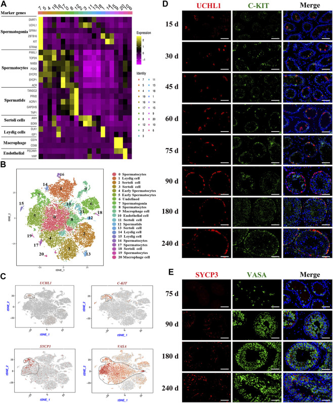
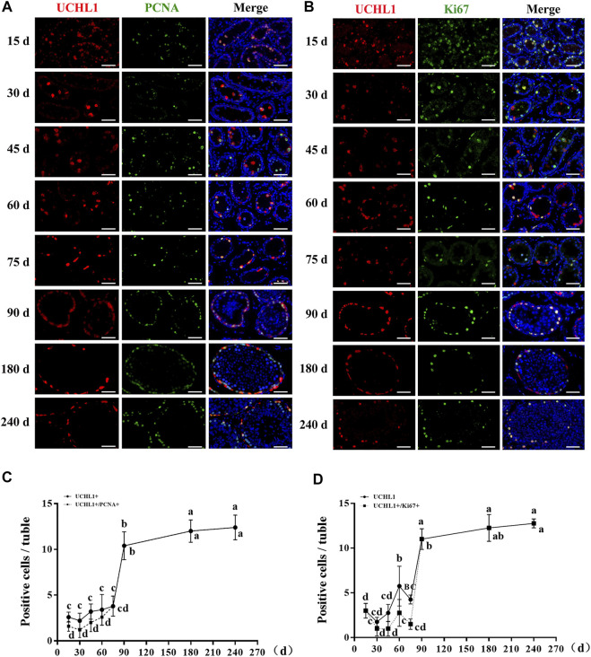
Spermatogenesis holds considerable promise for human-assisted reproduction and livestock breeding based on stem cells. It occurs in seminiferous tubules within the testis, which mainly comprise male germ cells and Sertoli cells. While the developmental progression of male germ cells and Sertoli cells has been widely reported in mice, much less is known in other large animal species, including dairy goats. In this study, we present the data of single cell RNA sequencing (scRNA-seq) for 25,373 cells from 45 (pre-puberty), 90 (puberty), and 180-day-old (post-puberty) dairy goat testes. We aimed to identify genes that are associated with key developmental events in male germ cells and Sertoli cells. We examined the development of spermatogenic cells and seminiferous tubules from 15, 30, 45, 60, 75, 90, 180, and 240-day-old buck goat testes. scRNA-seq clustering analysis of testicular cells from pre-puberty, puberty, and post-puberty goat testes revealed several cell types, including cell populations with characteristics of spermatogonia, early spermatocytes, spermatocytes, spermatids, Sertoli cells, Leydig cells, macrophages, and endothelial cells. We mapped the timeline for male germ cells development from spermatogonia to spermatids and identified gene signatures that define spermatogenic cell populations, such as AMH, SOHLH1, INHA, and ACTA2. Importantly, using immunofluorescence staining for different marker proteins (UCHL1, C-KIT, VASA, SOX9, AMH, and PCNA), we explored the proliferative activity and development of male germ cells and Sertoli cells. Moreover, we identified the expression patterns of potential key genes associated with the niche-related key pathways in male germ cells of dairy goats, including testosterone, retinoic acid, PDGF, FGF, and WNT pathways. In summary, our study systematically investigated the elaborate male germ cells and Sertoli cells developmental patterns in dairy goats that have so far remained largely unknown. This information represents a valuable resource for the establishment of goat male reproductive stem cells lines, induction of germ cell differentiation in vitro, and the exploration of sequential cell fate transition for spermatogenesis and testicular development at single-cell resolution.

Keywords: Sertoli cell; dairy goat; single-cell RNA sequencing (scRNA-seq); spermatogenesis; testes.


Conflict of interest statement

The authors declare that the research was conducted in the absence of any commercial or financial relationships that could be construed as a potential conflict of interest.

Figures

FIGURE 1
FIGURE 1
Overview of scRNA-seq in dairy goat testicular cells. (A) The single-cell RNA-seq process of dairy goat testicular cells. (B) Histomorphological analysis of dairy goat testes at different ages. 0, gonocyte. 1, spermatogonia. 2, early spermatocytes. 3, spermatocytes. 4, round spermatids. 5, elongated spermatids. 6, Sertoli cells. 7, Leydig cells. Bar = 100 μM. (C) Variation in the diameter of the seminiferous tubule of dairy goat testes. (D) Variation in the diameter of the lumen of the seminiferous tubule of dairy goat testes. (E) Single cell datasets quality metrics summary identified by Cell Ranger. (F) The reads mapped to genome of three samples. (G) Sample distribution result revealed in the tSNE plot. (H) Cell clustering revealed in the tSNE plot.
FIGURE 2
FIGURE 2
Identification of cell types during spermatogenesis and male germ cells development in dairy goat testes. (A) The marker genes specifically expressed of the cell cluster in heat map. (B) Cell clusters identification in the tSNE plot. (C) Expression patterns marker genes for male germ cells visualized in t-SNE plots. (D) UCHL1 and C-KIT immunostaining of dairy goat testis sections at each age. Bar = 100 μM. (E) SYCP3 and VASA immunostaining of dairy goat testis sections at 75 to 240-day-old. Bar = 100 μM.
FIGURE 3
FIGURE 3
The proliferative activity of spermatogonia in dairy goat testes. (A) UCHL1 and PCNA immunostaining of dairy goat testis sections at each age. Bar = 100 μM. (B) UCHL1 and Ki67 immunostaining of dairy goat testis sections at each age. Bar = 100 μM. (C) The numbers of UCHL1/PCNA-positive cells per cross-section of seminiferous tubules in dairy goat testes at each age (n = 9). (D) The numbers of UCHL1/Ki67-positive cells per cross-section of seminiferous tubules in dairy goat testes at each age (n = 9). Different letters imply significant difference (p < 0.05), and same letters mean no significant difference (p > 0.05).
FIGURE 4
FIGURE 4
Marker gene identification of each testicular cell type. (A) Heatmaps show the top 4 significantly differentially expressed genes between each cell cluster for testicular cells. (B,C) Expression patterns (violin plot) of each cell specific gene across the different cluster. (D) Immunofluorescence analysis the expression location of SOHLH1, INHA, and ACTA2 in dairy goat testes. Three potential markers (SOHLH1, spermatogonia; INHA, Sertoli cells; ACTA2, testicular somatic cells) and male germ cell marker (VASA) were stained in adult testis tissue sections, respectively. The DAPI (blue) denotes nuclei. SPG, spermatogonia. SC, Sertoli cell. LC, Leydig cell. PMC, peritubular myoid cells. Bar = 10 μm.
FIGURE 5
FIGURE 5
Construction of germ cell differentiation lineage in dairy goats and regulating gene expression patterns. (A) Cell trajectory analysis of nine testicular germ cell types. (B) Heat map of cell-cycle-specific genes in male dairy goat germ cells. (C) The indicator map of germ cell developmental changes. (D) Dot plot illustrating of germ cells key genes expressed in cell clusters. (E) Identification of cell clusters expressing the noted marker genes allowed clusters to be aligned with specific spermatogenic cell types.
FIGURE 6
FIGURE 6
Sertoli cells development in dairy goat testes. (A) Expression patterns AMH for Sertoli cells visualized in t-SNE plots. (B) Expression patterns (violin plot) of AMH the different cluster. (C) AMH and SOX9 immunostaining of dairy goat testis sections at each age. Bar = 100 μM. (D) The numbers of SOX9/PCNA-positive cells per cross-section of seminiferous tubules in dairy goat testes at each age (n = 9). Different letters imply significant difference (p < 0.05), and same letters mean no significant difference (p > 0.05). (E) SOX9 and PCNA immunostaining of dairy goat testis sections at each age. Bar = 100 μM.
FIGURE 7
FIGURE 7
Male germ cells and niche-related gene expression of key pathways in dairy goat. (A) Male germ cells and niche-related gene expression in the tSNE plot. (B) Heat map illustrating showed related genes in the male germ cells and Niche respectively.
FIGURE 8
FIGURE 8
Schematic diagram of postnatal male germ cells and Sertoli cells development dynamics in dairy goat.

Similar articles

Cited by

References

    1. Banco B., Palmieri C., Sironi G., Fantinato E., Veronesi M. C., Groppetti D., et al. (2016). Immunohistochemical expression of SOX9 protein in immature, mature, and neoplastic canine Sertoli cells. Theriogenology 85, 1408–1414. 10.1016/j.theriogenology.2015.12.024 - DOI - PubMed
    1. Cao C., Ma Q., Mo S., Shu G., Liu Q., Ye J., et al. (2021). Single-Cell RNA sequencing defines the regulation of spermatogenesis by Sertoli-Cell androgen signaling. Front. Cell. Dev. Biol. 9, 763267. 10.3389/fcell.2021.763267 - DOI - PMC - PubMed
    1. Chaichanathong S., Taya K., Watanabe G., Nagaoka K., Wajjwalku W., Sudsukh A., et al. (2018). Immunohistochemical localization of inhibin/activin subunits in adult Asian elephant (Elephas maximus) testes. J. Veterinary Med. Sci. 80, 549–552. 10.1292/jvms.17-0531 - DOI - PMC - PubMed
    1. Clark S., Mora García M. B. (2017). A 100-Year Review: Advances in goat milk research. J. Dairy Sci. 100, 10026–10044. 10.3168/jds.2017-13287 - DOI - PubMed
    1. De Oliveira C. S., Nixon B., Lord T. (2022). A scRNA-seq approach to identifying changes in spermatogonial stem cell gene expression following in vitro culture. Front. Cell. Dev. Biol. 10, 782996. 10.3389/fcell.2022.782996 - DOI - PMC - PubMed