

Human mesenchymal amniotic fluid stem cells reveal an unexpected neuronal potential differentiating into functional spinal motor neurons
- PMID: 35938174
- PMCID: PMC9354810
- DOI: 10.3389/fcell.2022.936990
Human mesenchymal amniotic fluid stem cells reveal an unexpected neuronal potential differentiating into functional spinal motor neurons
Abstract
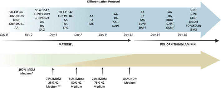
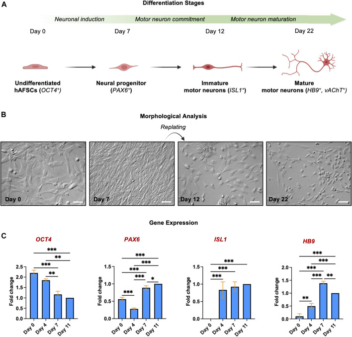
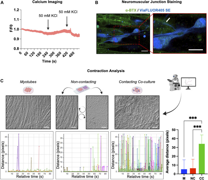
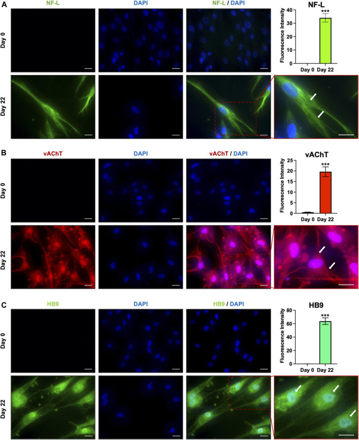
Human amniotic fluids stem cells (hAFSCs) can be easily isolated from the amniotic fluid during routinely scheduled amniocentesis. Unlike hiPSCs or hESC, they are neither tumorigenic nor immunogenic and their use does not rise ethical or safety issues: for these reasons they may represent a good candidate for the regenerative medicine. hAFSCs are generally considered multipotent and committed towards the mesodermal lineages; however, they express many pluripotent markers and share some epigenetic features with hiPSCs. Hence, we hypothesized that hAFSCs may overcome their mesodermal commitment differentiating into to ectodermal lineages. Here we demonstrated that by the sequential exposure to specific factors, hAFSCs can give rise to spinal motor neurons (MNs), as evidenced by the gradual gene and protein upregulation of early and late MN markers (PAX6, ISL1, HB9, NF-L, vAChT). When co-cultured with myotubes, hAFSCs-derived MNs were able to create functional neuromuscular junctions that induced robust skeletal muscle contractions. These data demonstrated the hAFSCs are not restricted to mesodermal commitment and can generate functional MNs thus outlining an ethically acceptable strategy for the study and treatment of the neurodegenerative diseases.
Keywords: amniotic fluid stem cells (AFSC); mesenchimal cells; motoneuron (MN); perinatal stem cells; regenerative medicine.
Copyright © 2022 Gaggi, Di Credico, Guarnieri, Mariggiò, Di Baldassarre and Ghinassi.
Conflict of interest statement
The authors declare that the research was conducted in the absence of any commercial or financial relationships that could be construed as a potential conflict of interest.
Figures
Similar articles
-
Human fetal membrane-mesenchymal stromal cells generate functional spinal motor neurons in vitro.iScience. 2022 Sep 23;25(10):105197. doi: 10.1016/j.isci.2022.105197. eCollection 2022 Oct 21. iScience. 2022. PMID: 36238899 Free PMC article.
-
The potential use of stem cells derived from human amniotic fluid in renal diseases.Kidney Int Suppl (2011). 2011 Sep;1(3):77-82. doi: 10.1038/kisup.2011.18. Kidney Int Suppl (2011). 2011. PMID: 25028628 Free PMC article. Review.
-
Recruitment of host's progenitor cells to sites of human amniotic fluid stem cells implantation.Biomaterials. 2011 Jun;32(18):4218-27. doi: 10.1016/j.biomaterials.2010.12.028. Epub 2011 Apr 2. Biomaterials. 2011. PMID: 21459439
-
Human Neuromuscular Junction on a Chip: Impact of Amniotic Fluid Stem Cell Extracellular Vesicles on Muscle Atrophy and NMJ Integrity.Int J Mol Sci. 2023 Mar 3;24(5):4944. doi: 10.3390/ijms24054944. Int J Mol Sci. 2023. PMID: 36902375 Free PMC article.
-
Amniotic fluid stem cells as a novel strategy for the treatment of fetal and neonatal neurological diseases.Placenta. 2021 Jan 15;104:247-252. doi: 10.1016/j.placenta.2021.01.009. Epub 2021 Jan 12. Placenta. 2021. PMID: 33461069 Review.
Cited by
-
Machine learning identifies phenotypic profile alterations of human dopaminergic neurons exposed to bisphenols and perfluoroalkyls.Sci Rep. 2023 Dec 11;13(1):21907. doi: 10.1038/s41598-023-49364-y. Sci Rep. 2023. PMID: 38081991 Free PMC article.
-
Impact on peri-implant connective tissue of laser treated versus traditional healing abutments: a human clinical trials.BMC Oral Health. 2023 Jun 27;23(1):425. doi: 10.1186/s12903-023-03148-y. BMC Oral Health. 2023. PMID: 37370064 Free PMC article.
-
Betaine Treatment Prevents TNF-α-Mediated Muscle Atrophy by Restoring Total Protein Synthesis Rate and Morphology in Cultured Myotubes.J Histochem Cytochem. 2023 Apr;71(4):199-209. doi: 10.1369/00221554231165326. Epub 2023 Apr 3. J Histochem Cytochem. 2023. PMID: 37013268 Free PMC article.
-
The Effects of Combined Exposure to Bisphenols and Perfluoroalkyls on Human Perinatal Stem Cells and the Potential Implications for Health Outcomes.Int J Mol Sci. 2023 Oct 9;24(19):15018. doi: 10.3390/ijms241915018. Int J Mol Sci. 2023. PMID: 37834465 Free PMC article.
-
Unraveling the Epigenetic Landscape: Insights into Parkinson's Disease, Amyotrophic Lateral Sclerosis, and Multiple Sclerosis.Brain Sci. 2024 May 29;14(6):553. doi: 10.3390/brainsci14060553. Brain Sci. 2024. PMID: 38928553 Free PMC article. Review.
References
-
- Antonucci I., Di Pietro R., Alfonsi M., Centurione M. A., Centurione L., Sancilio S., et al. (2014). Human second trimester amniotic fluid cells are able to create embryoid body-like structures in vitro and to show typical expression profiles of embryonic and primordial germ cells. Cell. Transpl. 23, 1501–1515. 10.3727/096368914X678553 - DOI - PubMed
LinkOut - more resources
Full Text Sources