Review
. 2022 Dec;247(23):2152-2172.
doi: 10.1177/15353702221110813. Epub 2022 Aug 6.

Recent advances in nanotechnology-enabled biosensors for detection of exosomes as new cancer liquid biopsy

Affiliations
Review

Recent advances in nanotechnology-enabled biosensors for detection of exosomes as new cancer liquid biopsy

Chang-Chieh Hsu et al. Exp Biol Med (Maywood). 2022 Dec.

Abstract

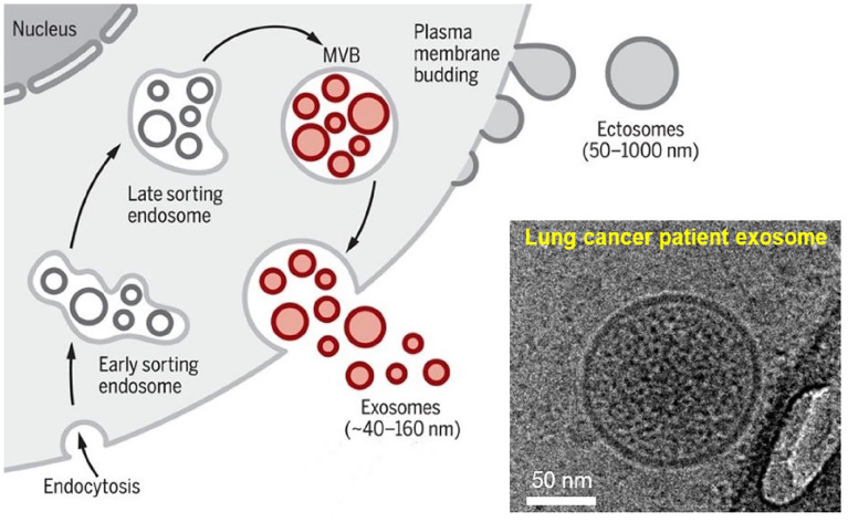
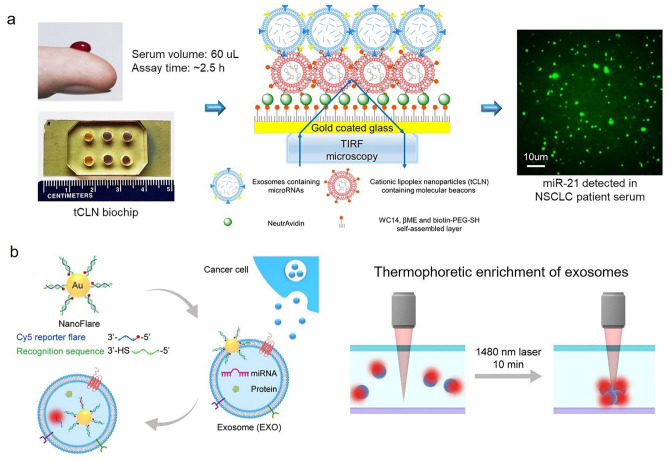
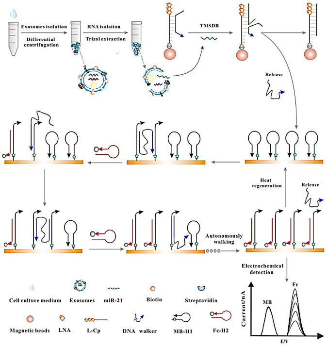
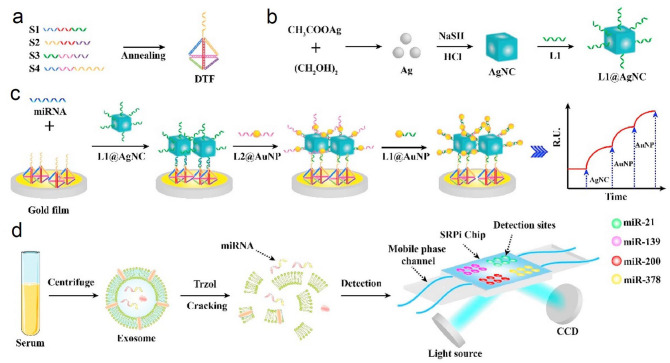
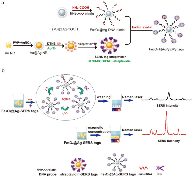
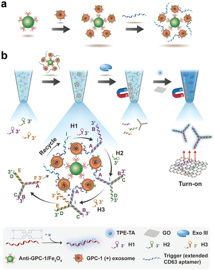
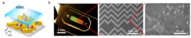
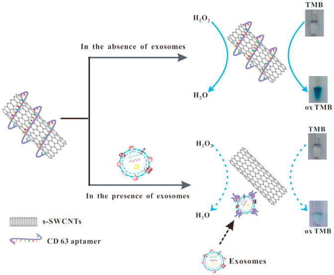
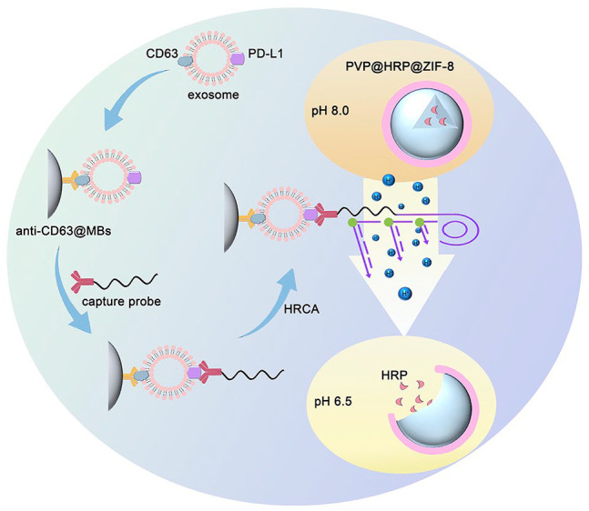
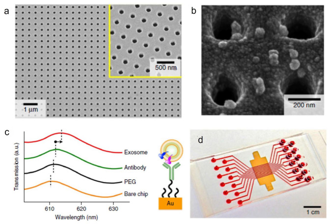
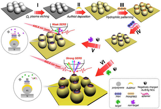
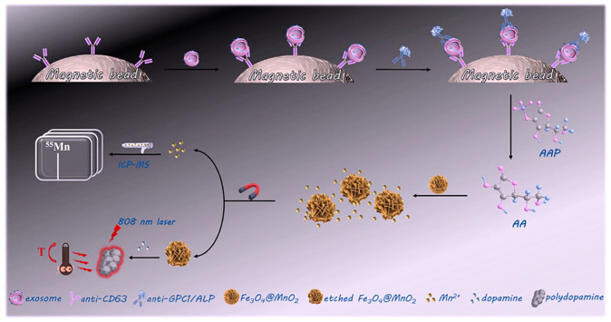
Cancer liquid biopsy detects circulating biomarkers in body fluids, provides information that complements medical imaging and tissue biopsy, allows sequential monitoring of cancer development, and, therefore, has shown great promise in cancer screening, diagnosis, and prognosis. Exosomes (also known as small extracellular vesicles) are cell-secreted, nanosized vesicles that transport biomolecules such as proteins and RNAs for intercellular communication. Exosomes are actively involved in cancer development and progression and have become promising circulating biomarkers for cancer liquid biopsy. Conventional exosome characterization methods such as quantitative reverse transcription polymerase chain reaction (qRT-PCR) and enzyme-linked immunosorbent assay (ELISA) are limited by low sensitivity, tedious process, large sample volume, and high cost. To overcome these challenges, new biosensors have been developed to offer sensitive, simple, fast, high throughput, low sample consumption, and cost-effective detection of exosomal biomarkers. In this review, we summarized recent advances in nanotechnology-enabled biosensors that detect exosomal RNAs (both microRNAs and mRNAs) and proteins for cancer screening, diagnosis, and prognosis. The biosensors were grouped based on their sensing mechanisms, including fluorescence-based biosensors, colorimetric biosensors, electrical/electrochemical biosensors, plasmonics-based biosensors, surface-enhanced Raman spectroscopy (SERS)-based biosensors, and inductively coupled plasma mass spectrometry (ICP-MS) and photothermal biosensors. The future directions for the development of exosome-based biosensors were discussed.

Keywords: Exosomes; biosensors; cancer liquid biopsy; extracellular vesicles; mRNA; microRNA; protein.


Conflict of interest statement

The author(s) declared no potential conflicts of interest with respect to the research, authorship, and/or publication of this article.

Figures

Figure 1.
Figure 1.
Exosome biogenesis and secretion. Exosomes are formed by inward budding of cell membrane to form intraluminal vesicles in early endosomes, which then develop into multivesicular bodies (MVB). MVB fuse with cell membrane and release exosomes. (Reprinted from Kalluri et al. ©2020 American Association for the Advancement of Science.) Inset: a CryoTEM image of an exosome in the blood of a lung cancer patient. (Reprinted from Wu et al. ©2013 American Chemical Society.) (A color version of this figure is available in the online journal.)
Figure 2.
Figure 2.
Sensing mechanisms of two representative fluorescence-based biosensors for exosomal RNA detection. (a) Sensing mechanism of tethered cationic lipoplex nanoparticles (tCLN) biochip in detecting exosomal RNAs. tCLN captured exosomes through electrostatic interaction. The fusion of exosomes with tCLN allowed the binding between molecular beacons and exosomal RNAs and thus restored the fluorescence signals from molecular beacons, which were detected by the total internal reflection fluorescence (TIRF) microscopy. (Reprinted from Liu et al. ©2020 Frontiers.) (b) Sensing mechanism of nanoflares-based thermophoretic biosensor in detecting exosomal RNAs. After nanoflares were internalized inside exosomes, the hybridization of recognition sequences with target microRNAs released Cy5 reporter flare and restored fluorescence signals. Then laser irradiation was applied to enable thermophoretic enrichment of exosomes to amplify fluorescence signals. (Reprinted from Zhao et al. ©2020 American Chemical Society.) (A color version of this figure is available in the online journal.)
Figure 3.
Figure 3.
Sensing mechanism of the DNA strand displacement reaction–based electrochemical biosensor in detecting exosomal miR-21 for breast cancer diagnosis. miR-21 was extracted from exosomes isolated from serum. Locked nucleic acid–modified capture probes (L-Cp) were conjugated on the surface of magnetic beads. DNA walkers partially hybridized to L-Cp to form DNA walker-L-Cp-magnetic bead complexes. In the presence of miR-21, L-Cp hybridized with miR-21 and the DNA walkers were released into the solutions. DNA walkers were then extracted using magnetic separation and applied to DNA tracks, which were made from gold electrodes. After DNA walkers bound to MB-H1 probes immobilized on the surface, Fc-H2 probes were added to hybridize with MB-H1 probes and initiate the walking of DNA walkers along the track. The approach of Fc-H2 probes to the surface led to the increase of electrochemical signals from Fc. The electrochemical signal ratio of Fc and MB was measured and used to characterize the expression of exosomal miR-21. (Reprinted from Zhang et al. ©2017 Elsevier B.V.) (A color version of this figure is available in the online journal.)
Figure 4.
Figure 4.
Sensing mechanism of a surface plasmon resonance imaging (SPRi) biosensor for multiplex detection of exosomal microRNAs. (a) A DNA tetrahedral framework (DTF) was prepared by self-assembly and attached to the surface of the SPRi biochip. (b) Single-strand DNA L1-modified silver nanocubes (L1@AgNC) were prepared. (c) MicroRNA and L1@AgNC are then applied onto the SPRi biochip to form DNA DTF-microRNA-AgNC complexes. Finally, single-strand DNA L2-modified gold nanoparticles (L2@AuNP) and single-strand DNA L1-modified gold nanoparticles (L1@AuNP) were sequentially added to form Au-on-Ag heterostructures, which amplified the SPR signals for sensitive detection of microRNA targets. (d) The workflow of SPRi assay. Exosomes were isolated from serum samples via centrifugation. Trizol was used to extract microRNAs from exosomes. The expression of microRNAs was measured by the SPRi assay. (Reprinted from Wu et al. ©2020 Elsevier B.V.) (A color version of this figure is available in the online journal.)
Figure 5.
Figure 5.
Sensing mechanism of dual-SERS biosensor for exosomal miR-10b detection for pancreatic cancer diagnosis. (a) Fe3O4@Ag-SERS tags were prepared by conjugating Fe3O4@Ag-DNA-biotin with SERS-tag-streptavidin (i.e. Au@Ag@DNTB-streptavidin). (b) In the presence of miR-10b and duplex-specific nuclease (DSN), SERS tags were continuously cleaved from Fe3O4@Ag and resulted in the reduction of SERS signals. (Reprinted from Pang et al. ©2019 Elsevier B.V.) (A color version of this figure is available in the online journal.)
Figure 6.
Figure 6.
Sensing mechanism of the hMFEX nanosensor. (a) Exosomes expressing GPC1 were captured by GPC1 antibody conjugated magnetic beads and subsequently bound with extended CD63 aptamers, forming the magnetic bead-exosome-aptamer complexes. (b) The complexes were mixed with three DNA hairpins to trigger a toehold-mediated strand displacement (TMSD) reaction among the DNA hairpins and produced DNA three-way junctions (TWJs). Tertiary amine-containing tetraphenylene (TPE-TA) and graphene oxide (GO) were added to generate fluorescence signals from DNA TWJs, allowing the quantification of exosomal GPC1 expression. (Reprinted from Li et al. ©2020 American Chemical Society.) (A color version of this figure is available in the online journal.)
Figure 7.
Figure 7.
Sensing mechanism of a 3D-nanopatterned herringbone (nano-HB) microfluidic device. (a) Schematic of the 3D-nano-HB microfluidic device. Porous herringbone structures offered large surface area for antibody immobilization to enhance exosome capture efficiency. The expression of exosomal proteins was quantified by fluorogenic ELISA. (b) A nano-HB device fabricated with 960 nm silica colloids. (Reprinted from Zhang et al. ©2019 Nature America, Inc.) (A color version of this figure is available in the online journal.)
Figure 8.
Figure 8.
Sensing mechanism of the colorimetric aptasensor for exosomal CD63 detection. CD63 aptamers absorbed on SWCNTs improved the peroxidase activity of SWCNTs, which catalyzed the oxidation of 3,3′,5,5′- tetramethylbenzidine (TMB) in the presence of H2O2 and changed the solution from colorless to deep blue. In the presence of exosomes, CD63 aptamers bound to exosomes and were released from the surface of SWCNTs. This process transformed the SWCNTs into their original state, decreased catalytic activity, and changed the color of the solution from deep blue to moderate blue. (Reprinted from Xia et al. ©2017 Elsevier B.V.) (A color version of this figure is available in the online journal.)
Figure 9.
Figure 9.
Schematic of detection of exosomal PD-L1 using a ZIF-8 MOF-based electrochemical biosensor for breast cancer diagnosis. Magnetic beads conjugated with CD63 antibodies were first used to capture exosomes. Then PD-L1 antibodies modified capture probes were added to bind to exosomes expressing PD-L1. The capture probes then served as the primers to initiate hyperbranched rolling circle amplification, which lowered the pH and led to the disassembly of PV@HRP@ZIF-8 MOFs and the release of HRP enzyme. The HRP enzyme generated electrochemical signals which were correlated with the expression of exosomal PD-L1. (Reprinted with permission from Cao et al. ©2020 Elsevier B.V.) (A color version of this figure is available in the online journal.)
Figure 10.
Figure 10.
Design and sensing mechanism of nPLEX biosensor. (a) An SEM image of periodic nanoholes in the nPLEX biosensor. (b) An SEM image of exosomes captured by antibodies modified on the surface of the nPLEX biosensor. (c) Representative responses (transmission spectra shifts and intensity increases) to the binding of PEG, antibody, and exosomes on the nPLEX biochip. (d) A picture of nPLEX biochip integrated with a multichannel microfluidic cell. (Reprinted from Im et al. ©2014 Nature America, Inc.) (A color version of this figure is available in the online journal.)
Figure 11.
Figure 11.
Sensing mechanism of SERS aptasensor based on a hydrophobic assembled nanoacorn (HANA) platform. The HANA platform was prepared by depositing a thin gold film on polystyrene nanosphere arrays, followed by layer-by-layer assembly of polyacrylic acid (PAA)/Rhodamine 6G (R6G), poly(allylamine hydrochloride) (PAH) and aptamers onto the surfaces of the plasmonic substrate to form ultrathin, hydrophilic sensing patches. SERS nanoprobes (Au@Ag nanocubes) were then added for exosomal protein detection. In the absence of exosomes, the negatively charged Au@Ag nanocubes were not able to bind on the sensing patches because of strong electrical repulsion. In the presence of exosomes, aptamers bound with exosomes and dissociated from the plasmonic substrate, which allowed the assembly of Au@Ag nanocubes on the sensing patches, forming a nanoparticle-on-mirror (NPoM) array. This NPoM array greatly enhanced SERS signals of Rhodamine 6G, enabling sensitive detection of exosomal proteins. (Reprinted from Zhu et al. ©2020 American Chemical Society.) (A color version of this figure is available in the online journal.)
Figure 12.
Figure 12.
Sensing mechanism of ICP-MS and photothermal dual-readout assay for the detection of exosomal GPC1 for pancreatic cancer diagnosis. Magnetic beads modified with CD63 antibodies captured exosomes. Then exosomes expressing GPC1 were labeled by GPC1 antibodies conjugated with alkaline phosphatase (ALP). l-ascorbic acid 2-phosphate (AAP) and Fe3O4@MnO2 nanoflowers were added to initiate the hydrolysis of AAP by ALP and the etching of Fe3O4@MnO2 nanoflowers to release Mn2+. After magnetic separation, the level of Mn2+ in supernatant was quantified by ICP-MS. The reduced Fe3O4@MnO2 nanoflowers were reacted with dopamine to generate polydopamine nanoparticles, which were irradiated by a near-infrared laser to generate photothermal signals. (Reprinted from Zhang et al. ©2021 American Chemical Society.) (A color version of this figure is available in the online journal.)

Similar articles

Cited by

References

    1. Ignatiadis M, Sledge GW, Jeffrey SS. Liquid biopsy enters the clinic—implementation issues and future challenges. Nat Rev Clin Oncol 2021; 18:297–312 - PubMed
    1. Siravegna G, Marsoni S, Siena S, Bardelli A. Integrating liquid biopsies into the management of cancer. Nat Rev Clin Oncol 2017;14:531–48 - PubMed
    1. Kalluri R, LeBleu VS. The biology, function, and biomedical applications of exosomes. Science 2020;367:eaau6977 - PMC - PubMed
    1. Möller A, Lobb RJ. The evolving translational potential of small extracellular vesicles in cancer. Nat Rev Cancer 2020;20:697–709 - PubMed
    1. LeBleu VS, Kalluri R. Exosomes as a multicomponent biomarker platform in cancer. Trends Cancer 2020;6:767–74 - PubMed

Publication types

Substances