

Enzyme-triggered- and tumor-targeted delivery with tunable, methacrylated poly(ethylene glycols) and hyaluronic acid hybrid nanogels
- PMID: 35938558
- PMCID: PMC9477489
- DOI: 10.1080/10717544.2022.2105443
Enzyme-triggered- and tumor-targeted delivery with tunable, methacrylated poly(ethylene glycols) and hyaluronic acid hybrid nanogels
Abstract
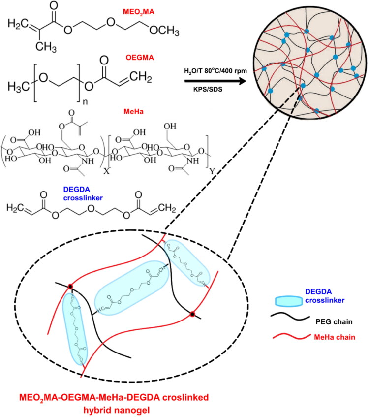
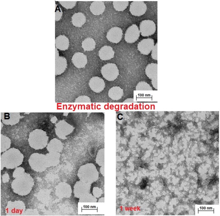
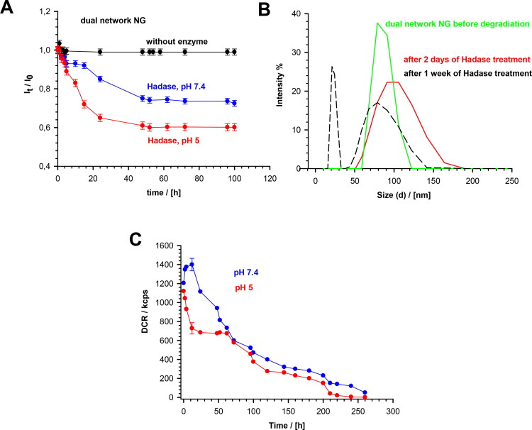
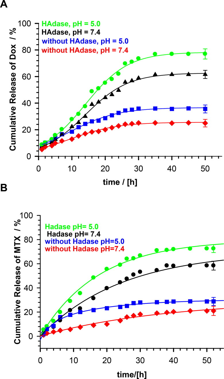
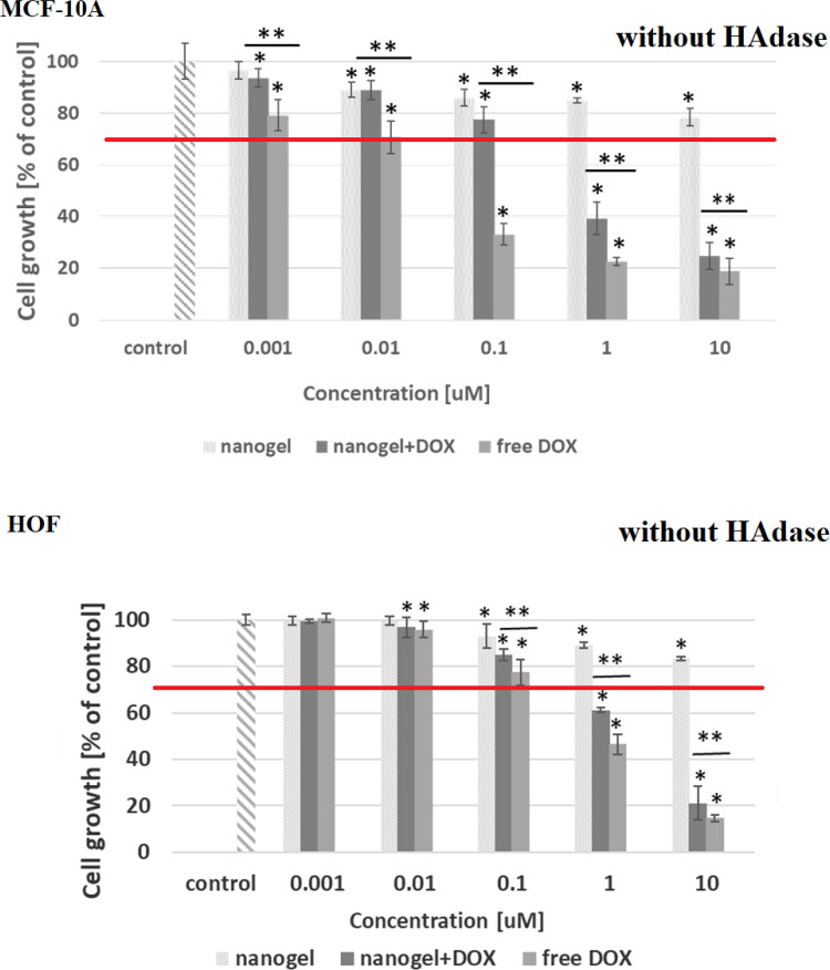
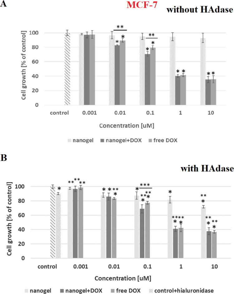
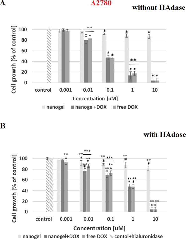
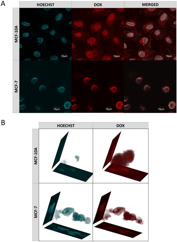
Enzyme-responsive polymeric-based nanostructures are potential candidates for serving as key materials in targeted drug delivery carriers. However, the major risk in their prolonged application is fast disassembling of the short-lived polymeric-based structures. Another disadvantage is the limited accessibility of the enzyme to the moieties that are located inside the network. Here, we report on a modified environmentally responsive and enzymatically cleavable nanogel carrier that contains a hybrid network. A properly adjusted volume phase transition (VPT) temperature allowed independent shrinking of a) poly(ethylene glycol) methyl ether methacrylate (OEGMA) with di(ethylene glycol) and b) methyl ether methacrylate (MEO2MA) part of the network, and the exposition of hyaluronic acid methacrylate (MeHa) network based carboxylic groups for its targeted action with the cellular based receptors. This effect was substantial after raising temperature in typical hyperthermia-based treatment therapies. Additionally, novel tunable NGs gained an opportunity to store- and to efficient-enzyme-triggered release relatively low but highly therapeutic doses of doxorubicin (DOX) and mitoxantrone (MTX). The controlled enzymatic degradation of NGs could be enhanced by introducing more hyaluronidase enzyme (HAdase), that is usually overexpressed in cancer environments. MTT assay results revealed effective cytotoxic activity of the NGs against the human MCF-7 breast cancer cells, the A278 ovarian cancer cells and also cytocompatibility against the MCF-10A and HOF healthy cells. The obtained tunable, hybrid network NGs might be used as a useful platform for programmed delivery of other pharmaceuticals and diagnostics in therapeutic applications.
Keywords: Targeted drug delivery; controlled release; enzymatic degradation; hybrid network nanogel; methacrylated hyaluronic acid; poly(ethylene glycol).
Conflict of interest statement
No potential conflict of interest was reported by the authors.
Figures
Similar articles
-
Doxorubicin Intracellular Remote Release from Biocompatible Oligo(ethylene glycol) Methyl Ether Methacrylate-Based Magnetic Nanogels Triggered by Magnetic Hyperthermia.ACS Appl Mater Interfaces. 2017 Aug 9;9(31):25775-25788. doi: 10.1021/acsami.7b06553. Epub 2017 Jul 27. ACS Appl Mater Interfaces. 2017. PMID: 28723064
-
Dual stimuli-responsive dendronized prodrug derived from poly(oligo-(ethylene glycol) methacrylate)-based copolymers for enhanced anti-cancer therapeutic effect.Acta Biomater. 2022 Apr 15;143:320-332. doi: 10.1016/j.actbio.2022.02.033. Epub 2022 Feb 27. Acta Biomater. 2022. PMID: 35235863
-
A novel dual-responsive core-crosslinked magnetic-gold nanogel for triggered drug release.Mater Sci Eng C Mater Biol Appl. 2016 Nov 1;68:436-444. doi: 10.1016/j.msec.2016.06.007. Epub 2016 Jun 5. Mater Sci Eng C Mater Biol Appl. 2016. PMID: 27524039
-
Bioresponsive functional nanogels as an emerging platform for cancer therapy.Expert Opin Drug Deliv. 2018 Jul;15(7):703-716. doi: 10.1080/17425247.2018.1497607. Epub 2018 Jul 16. Expert Opin Drug Deliv. 2018. PMID: 29976103 Review.
-
Smart stimuli sensitive nanogels in cancer drug delivery and imaging: a review.Curr Pharm Des. 2013;19(41):7203-18. doi: 10.2174/138161281941131219124142. Curr Pharm Des. 2013. PMID: 23489200 Review.
Cited by
-
Polymeric Gel Systems Cytotoxicity and Drug Release as Key Features for their Effective Application in Various Fields of Addressed Pharmaceuticals Delivery.Pharmaceutics. 2023 Mar 3;15(3):830. doi: 10.3390/pharmaceutics15030830. Pharmaceutics. 2023. PMID: 36986691 Free PMC article. Review.
-
Caspase-3-Responsive, Fluorogenic Bivalent Bottlebrush Polymers.ACS Macro Lett. 2024 May 21;13(5):571-576. doi: 10.1021/acsmacrolett.4c00119. Epub 2024 Apr 22. ACS Macro Lett. 2024. PMID: 38647178 Free PMC article.
-
Opportunities and Challenges of Switchable Materials for Pharmaceutical Use.Pharmaceutics. 2022 Oct 28;14(11):2331. doi: 10.3390/pharmaceutics14112331. Pharmaceutics. 2022. PMID: 36365149 Free PMC article. Review.
-
Degradable Nanogels Based on Poly[Oligo(Ethylene Glycol) Methacrylate] (POEGMA) Derivatives through Thermo-Induced Aggregation of Polymer Chain and Subsequent Chemical Crosslinking.Polymers (Basel). 2024 Apr 20;16(8):1163. doi: 10.3390/polym16081163. Polymers (Basel). 2024. PMID: 38675081 Free PMC article.
-
Ovarian cancer and its management through advanced drug delivery system.Med Oncol. 2025 Feb 17;42(3):76. doi: 10.1007/s12032-025-02621-8. Med Oncol. 2025. PMID: 39960609 Review.
References
-
- Amarsy I, Papot S, Gassera G. (2022). Stimuli-responsive metal complexes for biomedical applications. Angew. Chem. Int e202205900. - PubMed
-
- Amir MA, Khatoon F. (2019). Different types of smart nanogel for targeted delivery. J Sci Adv Mat Devices 4:201–12.
-
- Banerji S, Wright AJ, Noble M, et al. (2007). Structures of the Cd44–hyaluronan complex provide insight into a fundamental carbohydrate-protein interaction. Nat Struct Mol Biol 14:234–9. - PubMed
MeSH terms
Substances
LinkOut - more resources
Full Text Sources