

Biomimetic Tubular Matrix Induces Periodontal Ligament Principal Fiber Formation and Inhibits Osteogenic Differentiation of Periodontal Ligament Stem Cells
- PMID: 35938610
- PMCID: PMC10041666
- DOI: 10.1021/acsami.2c09420
Biomimetic Tubular Matrix Induces Periodontal Ligament Principal Fiber Formation and Inhibits Osteogenic Differentiation of Periodontal Ligament Stem Cells
Abstract
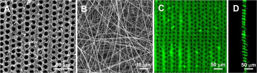
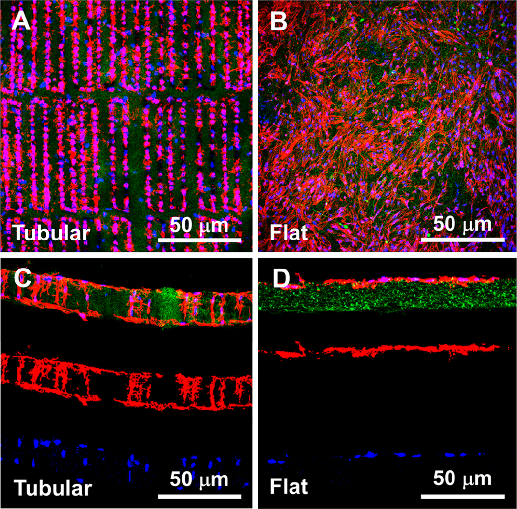
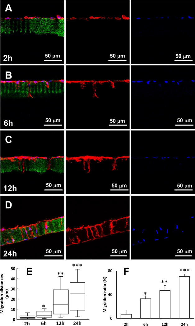
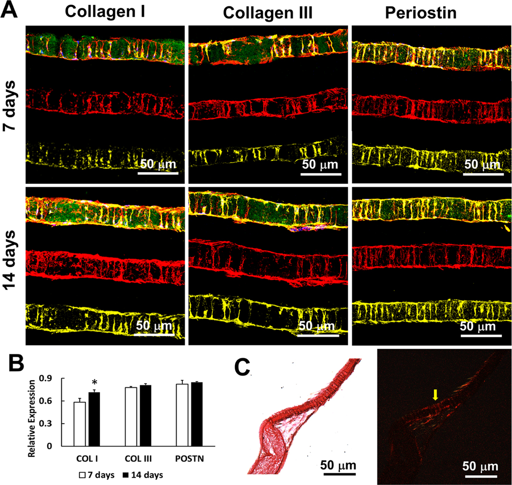
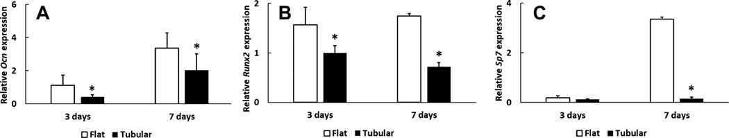
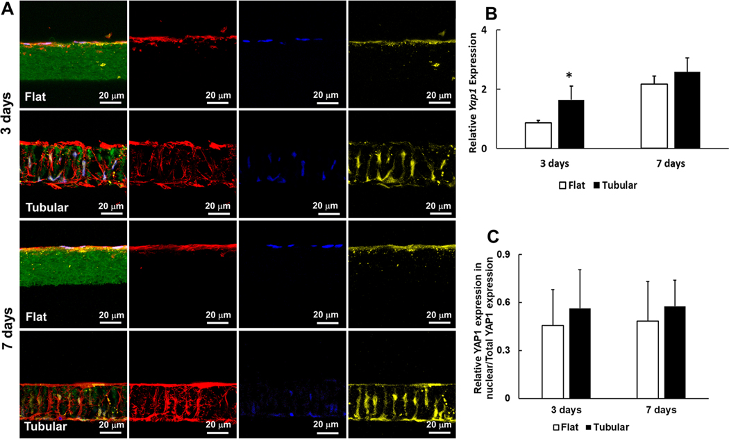
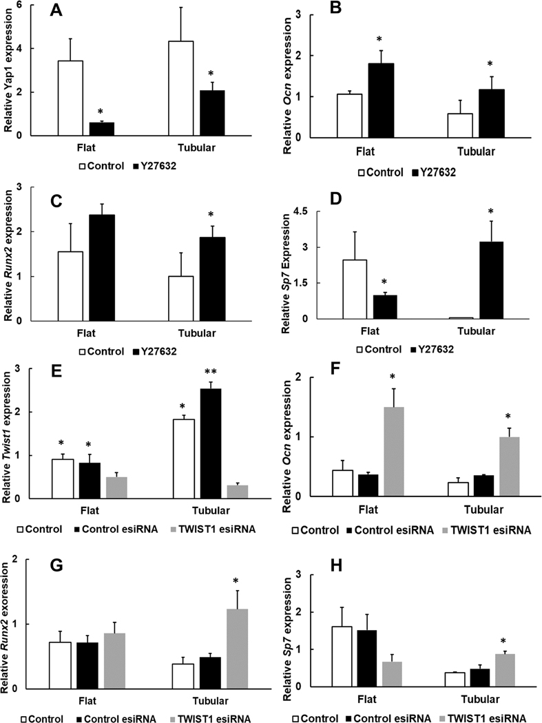
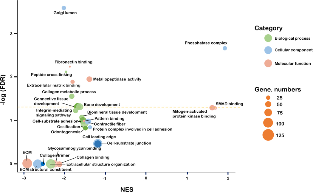
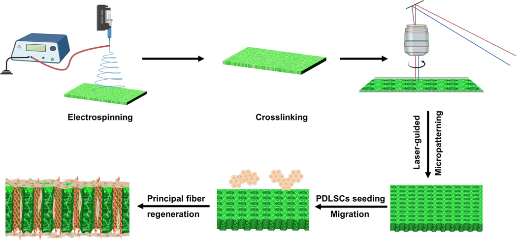
Periodontal ligament (PDL) is assembled from highly organized collagen fiber bundles (PDL principal fibers) that are crucial in supporting teeth and buffering mechanical force. Therefore, regeneration of PDL needs to reconstruct these well-ordered fiber bundles to restore PDL functions. However, the formation of PDL principal fibers has long been a challenge due to the absence of an effective three-dimensional (3D) matrix to guide the growth of periodontal ligament stem cells (PDLSCs) and to inhibit the osteogenic differentiation of PDLSCs during the PDL principal fibers deposition. In this work, we designed and fabricated a bio-inspired tubular 3D matrix to guide the migration and growth of human PDLSCs and form well-aligned PDL principal fibers. As a biomimetic 3D template, the tubular matrix controlled PDLSCs migration inside the tubules and aligned the cells to the designated direction. Inside the tubular matrix, the PDLSCs expressed PDL markers and formed oriented fiber bundles with the same size and density as those of natural PDL principal fibers. Furthermore, the tubular matrix downregulated the osteogenic differentiation of PDLSCs. A mechanism study revealed that the Yap1/Twist1 signaling pathway was involved in the inhibition of PDLSCs osteogenesis within the tubular matrix. This work provides an effective approach to induce PDLSCs to form principal fibers and gives insight into the underlying mechanism of inhibiting the osteogenic differentiation of PDLSCs in biomimetic tubular matrices.
Keywords: osteogenic differentiation; periodontal ligament; periodontal regeneration; principal fibers; tubular matrix.
Figures
Similar articles
-
A Novel Magnetic Manipulation Promotes Directional Growth of Periodontal Ligament Stem Cells.Tissue Eng Part A. 2023 Dec;29(23-24):620-632. doi: 10.1089/ten.TEA.2023.0112. Epub 2023 Sep 8. Tissue Eng Part A. 2023. PMID: 37603495
-
Characterization of stem cells from alveolar periodontal ligament.Tissue Eng Part A. 2011 Apr;17(7-8):1015-26. doi: 10.1089/ten.tea.2010.0140. Epub 2010 Dec 27. Tissue Eng Part A. 2011. PMID: 21186958
-
Pyrophosphate inhibits periodontal ligament stem cell differentiation and mineralization through MAPK signaling pathways.J Periodontal Res. 2021 Oct;56(5):982-990. doi: 10.1111/jre.12911. Epub 2021 Jun 17. J Periodontal Res. 2021. PMID: 34142719 Free PMC article.
-
Epigenetic regulation of osteogenic differentiation of periodontal ligament stem cells in periodontitis.Oral Dis. 2023 Oct;29(7):2529-2537. doi: 10.1111/odi.14491. Epub 2023 Jan 13. Oral Dis. 2023. PMID: 36582112 Review.
-
The relationship between MAPK signaling pathways and osteogenic differentiation of periodontal ligament stem cells: a literature review.PeerJ. 2025 Mar 31;13:e19193. doi: 10.7717/peerj.19193. eCollection 2025. PeerJ. 2025. PMID: 40183050 Free PMC article. Review.
Cited by
-
Nanomaterials Modulating the Fate of Dental-Derived Mesenchymal Stem Cells Involved in Oral Tissue Reconstruction: A Systematic Review.Int J Nanomedicine. 2023 Sep 21;18:5377-5406. doi: 10.2147/IJN.S418675. eCollection 2023. Int J Nanomedicine. 2023. PMID: 37753067 Free PMC article. Review.
-
Gli1+ Periodontal Mesenchymal Stem Cells in Periodontitis.J Dent Res. 2024 Mar;103(3):279-288. doi: 10.1177/00220345231220915. Epub 2024 Jan 29. J Dent Res. 2024. PMID: 38284236 Free PMC article.
-
Formulation of Hyperelastic Constitutive Model for Human Periodontal Ligament Based on Fiber Volume Fraction.Materials (Basel). 2025 Feb 6;18(3):705. doi: 10.3390/ma18030705. Materials (Basel). 2025. PMID: 39942371 Free PMC article.
-
Geometric Angles and Gene Expression in Cells for Structural Bone Regeneration.Adv Sci (Weinh). 2023 Nov;10(32):e2304111. doi: 10.1002/advs.202304111. Epub 2023 Sep 29. Adv Sci (Weinh). 2023. PMID: 37775309 Free PMC article.
-
Bioimplant-on-a-Chip for Facile Investigation of Periodontal Ligament Formation on Biogenic Hydroxyapatite/Ti6Al4 V Implants.ACS Appl Mater Interfaces. 2025 May 28;17(21):30673-30685. doi: 10.1021/acsami.5c04687. Epub 2025 May 13. ACS Appl Mater Interfaces. 2025. PMID: 40359253 Free PMC article.
References
-
- Papapanou PN; Susin C. Periodontitis Epidemiology: Is Periodontitis Under-Recognized, Over-Diagnosed, or Both? Periodontol. 2000 2017, 75, 45–51. - PubMed
-
- Slots J. Periodontitis: Facts, Fallacies and the Future. Periodontol. 2000 2017, 75, 7–23. - PubMed
-
- Tsumanuma Y; Iwata T; Washio K; Yoshida T; Yamada A; Takagi R; Ohno T; Lin K; Yamato M; Ishikawa I; Okano T; Izumi Y. Comparison of Different Tissue-Derived Stem Cell Sheets for Periodontal Regeneration in a Canine 1-Wall Defect Model. Biomaterials 2011, 32, 5819–5825. - PubMed
MeSH terms
Grants and funding
LinkOut - more resources
Full Text Sources
Research Materials
